
Assessing the severity of alopecia areata
INESSA SERBINA
Scientific consultant, TRICHOLOGY INSTITUTE ®
Сербина И. М. Оценка степени тяжести гнездной алопеции / И. М. Сербина // Дерматологія та венерологія. - 2014. - № 4. - С. 65-71.
Alopecia areata (AA) is currently regarded as a tissue-specific autoimmune disease mediated by autoactivated T lymphocytes directed at hair follicles in the anagen stage, in the setting of a breakdown of immune tolerance. The manifestation and clinical diversity of AA are determined by a complex combination of environmental and genetic factors [2,7,11].
The course of the disease is unpredictable. It may affect the scalp and/or hair on other areas, and may appear as isolated patches of hair loss or as extensive, rapid, complete hair loss. Hair may regrow spontaneously several months or years after the onset of the disease, or the hair loss may persist permanently despite therapeutic interventions [4,8]. In 60% of AA patients the first episode of hair loss occurs before the age of 20, and the incidence peaks between 20 and 40 years. In recent years there has been a trend towards rising incidence, especially among young people, and a high frequency of severe forms combined with various psycho-emotional disorders (asthenic symptoms, sleep and appetite disturbances, depression, etc.) and a marked reduction in patients' quality of life. For this reason, the development of effective treatments for AA remains a subject of active research by many scientists [2,7].
It is known that one of the main criteria for choosing treatment tactics for an AA patient is the severity of the dermatosis [1,8]. Today dermatologists worldwide use a large number of different indices and scales to characterise the severity of the condition of dermatological patients. This makes it possible to increase the accuracy of clinical assessments and to analyse the effectiveness of therapy. The use of an objective, unified scale for assessing severity in AA is relevant [6,9].
Most physicians in our country assess the severity of hair loss in AA using the classification of I.V. Shutsky [5], which distinguishes 4 degrees of severity: degree I – single lesions 3–5 cm in diameter; degree II – lesions 5–10 cm in diameter; degree III – subtotal alopecia; degree IV – total alopecia (TA) and universal alopecia (UA).
An analysis of the clinical picture of AA at the present stage shows that involvement of the scalp is often combined with hair loss on other areas of the skin, and is observed not only in UA, where it is considered to be a regular feature [7,12]. Dystrophy of the nail plates is a frequent symptom, and its presence is regarded as a prognostically unfavourable sign in the course of AA [9]. The existing classification does not take these factors into account.
An international group of experts in hair research, acting on behalf of the National Alopecia Areata Foundation, has developed a special standardised system, SBN, which makes it possible to objectify the assessment of hair loss on the scalp – S (Scalp), on the body – B (Body), and of involvement of the nail plates – N (Nail), as well as to monitor the effectiveness of treatment [3,6].
Degree of hair loss on the scalp (S0–S5):
• S0 – no hair loss;
• S1 – loss of <25% of hair;
• S2 – loss of 25 to 49% of hair;
• S3 – loss of 50 to 74% of hair;
• S4 – loss of 75–99 % of hair, specified as:
- S4а – loss of 75 to 95% of hair;
- S4b – loss of 96–99% of hair;
• S5 – loss of 100% of scalp hair.
Degree of hair loss on other areas of the body (В0-В2):
• В0 – hair preserved;
• В1 – partial hair loss;
• В2 – 100% hair loss.
Degree of nail plate changes (N0-N1а):• N0 – no involvement;
• N1 – partial involvement;
• N1а – dystrophy of all 20 nails.
E. Olsn and J. Canfield introduced the alopecia severity measure SALT (Severity of Alopecia Tool), which provides a formal mathematical approach to determining hair loss and hair regrowth [3,6,8]. Fig. 1 is used as a visual aid for assessing the degree of hair loss on the scalp.
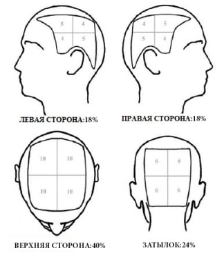
The entire surface of the scalp is taken as 100% and is conventionally divided into 4 quadrants, corresponding to the left, right, posterior and top sides of the scalp. The area of the left and right sides is 18% each; the area of the top (parietal and vertex regions) is 40%; the posterior side (occipital region) is 24%. Each quadrant is in turn divided into 4 parts, and the figures in them indicate what percentage the hair loss in that particular area makes up. The degree of hair loss in a patient with AA is assessed visually on all 4 sides of the scalp, in the corresponding zone of the quadrant, and the results are summed, that is, the total percentage of scalp hair loss is calculated. These data make it possible to determine the degree of hair loss on the head more precisely, according to the S0–S5 standards of the AA severity scale.
Using the SBN scale, the clinical variant of an AA patient with a multifocal form and a scalp involvement of 65%, with loss of eyelashes but without hair loss on other areas of the body and without nail damage, is expressed as S3B1N0.
The aim of our study was to examine the clinical features and to determine the degree of severity in AA patients using the standardised SBN assessment system.
Materials and methodsSixty patients with AA were under observation. By sex, the patients were distributed as follows: 22 men (36.6%) and 38 women (63.3%). The mean age of the patients was 30.4 years, in a range of 18-49 years. The data show that all patients were of active working age, with women predominating.
The diagnosis was established on the basis of the clinical picture and the findings of instrumental diagnostic examination. The methods of clinical and laboratory diagnosis followed a single plan, which included a general examination of the patient, standard laboratory tests (complete blood count, urinalysis, blood chemistry), and, as indicated, examination of the thyroid gland, consultation with related specialists, dermatotrichoscopy and a scalp skin biopsy.
Results and discussionThe onset of AA varied over a wide age range, from 5 to 45 years. The mean age at the start of the pathological process was 19.2±5.4 years. In 29.4% of patients with scalp hair loss over 25%, together with hair loss on other areas of the body and changes in the nail plates, the onset of AA was recorded before the age of 10. An analysis of disease onset in patients with scalp hair loss under 25% showed that in only 3.2% of the patients in this group did AA begin in the same age period.
The mean duration of the disease was 10.2 months, with a range from 2 weeks to 6 years. The number of relapses ranged from 1 to 8. More than 2 relapses occurred in 64.5% of patients with scalp hair loss over 25% and in only 12.4% of cases of the focal form of AA.
On external examination, the pattern of hair loss was determined in AA patients. The following clinical forms were diagnosed: focal (18 patients), band-like (ophiasis, inverse ophiasis) (12 patients), multifocal (17 patients), and total and universal (13 patients).
The focal form was represented by nummular patches on the scalp or, in men, the beard. The band-like form (ophiasis) was characterised by patches of hair loss in the occipital area which merge and spread in a band towards the auricles, reaching the temples. Inverse ophiasis (sisapho), a pattern of hair loss in the direction opposite to ophiasis (the frontal-temporal-parietal area), was found in 2 patients. The multifocal form was characterised by extensive areas of involvement resulting from the merging of smaller patches. TA was characterised by a complete absence of hair on the scalp. In UA, the bristly hair (eyebrows and eyelashes) was also absent, and there was partial or complete loss of vellus hair on the body.
According to the SBN scale, the hair loss state S1 corresponded clinically to focal alopecia, S2–S4 to ophiasic and multifocal alopecia, and S5 to TA and UA.
A full examination of the hair cover on the face, trunk, limbs, genital area and axillary zones was carried out in all forms of AA. Partial or complete hair loss in these areas was noted in 38% of patients with S2–S3 and in 67% with the S4–S5 clinical forms. When examining an AA patient, the condition of the nail plates was assessed. Various changes, such as pitting, longitudinal ridging, onychorrhexis, leukonychia and trachyonychia, were found in 19% of patients with S2–S3 and in 48% with S4–S5. A combination of varying degrees of body hair loss and nail plate dystrophy was noted in 26% of the AA patients examined.
The distribution of AA patients according to the SBN scale in our study is shown in Table 1.
Table 1
Distribution of patients with alopecia areata
| Clinical form of AA (SBN) | Number of patients |
| S1 В0 N0 | 18 |
| S2 В0 N0 | 6 |
| S2 В0 N1 | 1 |
| S2 B1 N0 | 2 |
| S2 В2 N0 | 1 |
| S2 В1 N1 | 1 |
| S3 В0 N0 | 6 |
| S3 В1 N0 | 2 |
| S3 В1 N1 | 2 |
| S4аB0 N0 | 1 |
| S4аB0 N1 | 3 |
| S4вB1 N0 | 2 |
| S4вB1 N1 | 2 |
| S5 B0 N0 | 3 |
| S5 B1 N0 | 1 |
| S5 B1 N1 | 2 |
| S5 B2 N0 | 4 |
| S5 B2 N1 | 2 |
| S5 B1 N1а | 1 |
| Total | 60 |
The analysis of the results showed that no additional clinical symptoms were observed in patients with hair loss under 25% (S1). Among patients with hair loss of 25-49% (S2), partial body hair loss was found in 2 (3.3%) and a change in the nail plates in 1 (1.7%), with both signs together in 2 (3.3%). Complete body hair loss was noted in 1 (1.7%) patient with the ophiasic form of the disease. Scalp hair loss of 50 to 74% (S3) was accompanied by hair loss on other areas in 2 (3.3%) and by a combination of body hair involvement and onychodystrophy in 3 (5%) of those examined. With scalp hair loss of 75 to 99% (S4), partial body hair loss was found in 2 patients (3.3%) and a change in the nail plates in 3 (5%), while 2 (3.3%) patients with S4в showed a combination of these signs. In patients with TA and UA, hair on other areas was completely absent in 4 (6.6%), and a combination of this sign with a change in the nail plates was observed in 2 (3.3%) patients. One patient with S5 had involvement of all nails of the trachyonychia type.
Thus, body hair loss and involvement of the nail plates were diagnosed when more than 25% of scalp hair was lost, and their frequency increased as the area of scalp involvement widened. Early onset of the disease and a high frequency of relapses were also noted more often in S2–S5 patients, which is regarded as an unfavourable prognostic criterion.
Since patches of alopecia on areas other than the scalp, as well as dystrophic changes of the nail plates, indicate the spread and worsening of the pathological process, the clinical variant S1В0N0 is regarded as a mild form, S2B0-2N0-1 – S3B0-2N0-1 as characterising moderate severity, and S4B0-2N0-1a–S5B0-2N0-1a as severe AA. The clinical heterogeneity of AA is important to take into account when determining treatment tactics [7,9]. Systemic treatment methods can be used even when scalp hair loss is 25-75%, if there is hair loss on other areas of the skin and/or nail dystrophy. The development of a scoring system for determining severity, combining a quantitative assessment of the severity of clinical manifestations with the area of scalp involvement, is promising.
ConclusionsIn assessing the severity of AA, what matters is not only the number and size of patches on the scalp but also the overall pattern of hair loss on the skin and the possible combination of hair loss with dystrophic changes of the nail plates, which are regarded as harbingers of a severe course of the disease. Classifying the severity of AA by the SBN system makes it possible to assess and describe the clinical variant according to a standardised scale, to determine treatment tactics for patients with various manifestations, and to predict the course of the disease.
References
1. Борисова О.Б. Лікування хворих на гніздову алопецію залежно від стадії та сту- пеня тяжкості захворювання / О.Б. Борисова // Дерматол. та венерол. – 2009. – No 3. – С.10–14.
2. Верхогляд И.В. Современные пред- ставления об этиологии, патогенезе и лече- нии очаговой алопеции / И.В. Верхогляд,О.Ю. Олисова, И.П. Гастроверхова // Россий- ский журнал кожных и венерических болез- ней. – 2010. – No 1. – С. 48–51.
3. Гаджигороева А.Г. Клиническая три- хология / А.Г. Гаджигороева. – М. : Практи- ческая медицина, 2014. – 184 с.
4. Исследование местных иммунных механизмов воспаления при гнездной ало- пеции с учётом активности заболевания / Н.Н. Потекаев, Е.А. Коган, А.Г. Гаджигоро- ева и др. // Клин. дерматология и венероло- гия. – 2010. – No 5. – С. 103–108.
5. Мавров И.И. Основы диагностики и лечения в дерматологии и венерологи: руко- водство для врачей, интернов, студентов / И.И. Мавров, Л.А. Болотная, И.М. Сербина. – Харьков: Факт, 2007. – 792 с.
6. Alopecia areata investigational assess- ment guidelines. Part II / E. Olsen, M. Hordin- sky, V. Price et al. // J. Am. Acad. Dermatol. – 2004. – Vol. 51. – Р. 440-447.
7. Alopecia areata update: part I. Clini- cal picture, histopathology, and pathogenesis / A. Alkhalifah, A. Alsantali, E. Wang et al. // J. Am. Acad. Dermatol. – 2010. – Vol. 62. – P. 177-188.
8. Al-Salam F.A. Alopecia areata: Updates / Al-Salam F.A., A.A. Azim, H. Darwish // The Gulf Journal of Dermatology and Venereology. – 2013. – Vol. 20, No. 2. – P. 1-16.
9. Finner A.M. Alopecia areata: Clinical presentation, diagnosis, and unusual cases / A.M. Finner // Dermatol. Ther. – 2011. – Vol. 24. – P. 348-354.
10. Investigation of the inflammatory mech- anisms in alopecia areata / E.D. Cetin, E. Savk, M. Uslu et al. // Am. J. Dermatopathol. – 2009. – Vol. 31, No. 1. – P. 53-60.
11. Ito Т. Recent Advances in the Pathogen- esis of Autoimmune Hair Loss Disease Alope- cia Areata / T. Ito // Clinical and Developmental Immunology. – 2013. – Article ID 348546. – P. 1-6.
12. Kasumagic-Halilovic E. Nail changes in alopecia areata: frequency and clinical presen- tation / E. Kasumagic-Halilovic, A. Prohic // J. Eur. Acad. Dermatol. Venereol. – 2009. – Vol. 23, No. 2. – P. 240-241.
References
1. Borysova O.B. Likuvannia khvorykh na hnizdovu alopetsiiu zalezhno vid stadii ta stu- penia tiazhkosti zakhvoriuvannia // Dermatol. ta venerol. – 2009. – N 3. – S.10-14.
2. Verhoglyad I.V,. Olisova O.Yu., Gastro- verhova I.P. Sovremennyie predstavleniya ob etiologii, patogeneze i lechenii ochagovoy alo- petsii / I.V. Verhoglyad, // Rossiyskiy zhurnal kozhnyih i venericheskih bolezney. – 2010. – N 1. – S. 48-51.
3. Gadzhigoroeva A.G. Klinicheskaya tri- hologiya. – M. : Prakticheskaya meditsina, 2014. – 184 s.
4. Potekaev N.N., Kogan E.A., Gadzhigoro- eva A.G. i dr. Issledovanie mestnyih immunnyih mehanizmov vospaleniya pri gnezdnoy alopet- sii s uchYotom aktivnosti zabolevaniya // Klin. dermatologiya i venerologiya. – 2010. – N
5. – S. 103-108. 5. Mavrov I.I Osnovyi diagnostiki i lech- eniya v dermatologii i venerologi: rukovodstvo dlya vrachey, internov, studentov – Harkov: Fakt, 2007. – 792 s.
6. Olsen E., Hordinsky M., Price V. et al. Alopecia areata investigational assessment guidelines. Part II // J. Am. Acad. Dermatol. – 2004. – Vol. 51. – Р. 440-447.
7. Alkhalifah A., Alsantali A., Wang E. et al. Alopecia areata update: part I. Clinical picture, histopathology, and pathogenesis // J.Am.Acad. Dermatol. – 2010. – Vol. 62. – P. 177-188.
8. Al-Salam F.A., Azim A.A., Darwish H. Alopecia areata: Updates // The Gulf Journal of Dermatology and Venereology. – 2013. – Vol. 20, No. 2. – P. 1-16.
9. Finner A.M. Alopecia areata: Clinical presentation, diagnosis, and unusual cases // Dermatol. Ther. – 2011. – Vol. 24. – P. 348-354.
10. Cetin E.D., Savk E., Uslu M. et al. Inves- tigation of the inflammatory mechanisms in alo- pecia areata // Am. J. Dermatopathol. – 2009. – Vol. 31, No. 1. – P. 53-60.
11. Ito Т. Recent Advances in the Pathogen- esis of Autoimmune Hair Loss Disease Alopecia Areata // Clinical and Developmental Immunol- ogy. – 2013. – Article ID 348546. – P. 1-6.
12. Kasumagic-Halilovic E., Prohic A. Nail changes in alopecia areata: frequency and clinical presentation // J. Eur. Acad. Dermatol. Venereol. – 2009. – Vol. 23, No. 2. – P. 240-241.