
Immunomorphological features of the development of alopecia areata
Scientific consultant, TRICHOLOGY INSTITUTE ®
Сербiна, I. М. Iмуноморфологічні особливості формування гніздової алопеції // ДЕРМАТОЛОГІЯ ТА ВЕНЕРОЛОГІЯ —№ 3 (73) — 2016 стр. 52-60
Introduction
The data obtained to date on the mechanisms of alopecia areata (AA) indicate that the pathogenesis of this disease is based on cell-mediated local immune reactions arising from a breakdown of the immune tolerance of hair follicles (HF), driven by genetic factors and exogenous triggers [4, 6, 10]. Histological and immunohistochemical (IHC) studies show a local tissue inflammatory reaction around the HF, and the peri- and intrafollicular cellular responses point to an immune attack against an unknown hair follicle antigen [5, 7]. T lymphocytes, namely CD4+ and CD8+, make up the bulk of the cells of the inflammatory infiltrate and obviously play a key role in the pathogenesis of AA. Under the influence of activated T helper cells, production of proinflammatory cytokines rises, together with a cascade of further immunopathological processes that lead to nonspecific autoimmune inflammation [1, 2, 11, 12]. Genome-wide association studies show that at a certain stage of AA, proinflammatory factors and ligands that stimulate NK cells and induce the collapse of HF immune privilege may become active [13, 15]. The literature describing the pathomorphological picture of AA is contradictory and does not fully cover the distribution of infiltrate cells and their immune phenotype, because it does not take the phase of the disease into account [8, 9, 11, 12].
The aim of our study was therefore to examine the morphological and IHC features of the acute and chronic stages of AA and to refine the pathogenesis of the disease on the basis of the data obtained. The tasks of the study included determining the distribution of CD4+, CD8+, CD68+ and CD56+ cells and the profiles of the proinflammatory cytokines IL-1, IL-6 and TNF-α in AA patches in the acute and chronic stages of the disease.
Materials and methodsScalp skin biopsies from 14 patients with a clinical diagnosis of AA, confirmed histologically and dermoscopically, were studied. The study included 8 women and 6 men aged 20 to 45 years. Skin biopsies from 5 healthy people, taken from the scalp during plastic surgery, were used as controls. The study was conducted after the patients signed informed consent in accordance with the regulations governing medical research.
According to the literature, the histological parameters of AA do not correlate with clinical and anamnestic data such as the number and size of patches or concomitant pathology, but depend only on the duration of the disease and the activity of the pathological process [11, 14]. For this reason, we also took into account only the clinical and dermoscopic parameters of AA activity and its duration. Seven patients had an active stage, with a duration of the disease or of the current episode averaging from 2-3 weeks to 6 months. The chronic stage, lasting from 6 months to 6 years or more, was found in the remaining 7 patients. None of the patients had received treatment for AA for 6 months or longer.
Skin samples from the areas of hair loss were taken with a disposable 4 mm punch biopsy. After the biopsy, the material was fixed in 4% formalin solution for 48 hours and embedded in paraffin. Serial sections 4-5 μm thick were prepared from the paraffin blocks. For routine staining, the histological sections were stained with haematoxylin and eosin. IHC examination was carried out by a standardised method on serial paraffin sections mounted on polylysine-coated adhesive slides («Menzel-Glaser», Germany), using DAKO reagents. The IHC panel included the following antibodies: CD4 (Clone 4B12), CD8 (Clone С8/144В), CD68 (Clone PG-M1), CD56 (Clone 123C3). Proinflammatory cytokines: TNF-α, IL-1, IL-6. Visualisation system EnVisionTM FLEX+, Mouse, High pH (Link), Code K8012 on a DAKO autostainer.
The intensity of cytoplasmic expression of CD68, CD56, TNF-α, IL-1 and IL-6 was assessed by a semiquantitative method based on staining intensity, using the following scheme: weak, moderate and strong expression. CD4 and CD8 – membrane reaction. Morphometric study of the markers included counting positive cells in 5 fields of view at magnification 200 using the DP SOFT program. The slides were viewed and digitally photographed with an «Olympus CX-4» light microscope.
Dermoscopic examination was carried out with a computer diagnostic program and Aramo SG videodermoscopy equipment (Korea), which gives images at magnifications of 60 and 200. The examination included determining the main dermoscopic signs of AA (yellow dots, black dots, dystrophic exclamation-mark hairs, vellus hairs) [4].
Results and discussion
Patients in the active stage of the disease complained of increased hair loss accompanied by the formation of patches of baldness; subjectively they felt moderate itching, burning and a sensation of «crawling ants» on the scalp, where alopecia patches formed later. On clinical examination the following were noted: patches of baldness with clear borders and signs of hyperaemia, with hairs broken off at a height of 1-2 mm in the patches and along their edge; on the hair pull test at the border of the patches there was a zone of loosened hair about 0.3-1 cm wide, in which telogen or dystrophic hairs were easily epilated. The chronic stage of AA was characterised by absence of hair growth and absence of subjective complaints related to impaired or altered skin sensation in the patches of baldness. The clinical picture of the disease had few symptoms: the skin in the patches was of normal colour and slightly hypotrophic, the follicular openings were not visualised, and there was no zone of loosened hair.
On dermoscopy, the number of yellow dots was highest in patients with the active stage of AA (24.3±7.2) and fell significantly, by a factor of 1.4, in the chronic stage (16.5±7.3). Dystrophic exclamation-mark hairs (4.9±0.8) and black dots (3.5±0.7) were present only while the pathological process was active. Vellus hairs, by contrast, were absent in patients with a progressive course of the dermatosis and appeared in the chronic stage (2.8±0.2).
In analysing the histological picture of the scalp skin in AA, attention was paid to the epidermis, the dermis and the HF, and the microcirculatory bed. The general morphological processes in AA were assessed, such as the presence and distribution of the inflammatory infiltrate, the presence of sclerosis, changes in the follicular epithelium, and the presence of hair in the HF. The structural changes at different stages of AA were found to be heterogeneous and to consist of inflammatory and dystrophic processes of varying severity.
In the active stage of AA, the findings were an unevenly thickened epidermis, especially near the superficial openings of the HF, acanthosis, moderate hyperkeratosis and thickening of the granular layer to 2-3 rows, isolated vacuolated epithelial cells, moderate intercellular oedema and hyperchromia of the nuclei of basal cells. In the dermis under the epidermis there was focal moderate lymphohistiocytic infiltration with an admixture of neutrophils, loosening of the collagen fibres, a reduced number of capillaries and increased permeability of the vessel wall, accompanied by perivascular oedema.
The patches contained small HF with a deformed hair bulb, corresponding to anagen phase IV or to the catagen, kenogen and telogen stages. In most HF the hair was absent. Multiple fragments of hair shafts were seen in anagen follicles. In skin biopsies taken in the zone of loosened hair, the openings of the hair follicles were dilated. However, dystrophic terminal hairs in the anagen phase were also found, surrounded by an inflammatory infiltrate. The outer and inner root sheaths showed structural disturbances with detachment of the connective tissue sheath, in which marked degenerative changes were noted. The epithelium of the hair bulb showed signs of proliferation, dys- and parakeratosis, and a few cells with intracellular oedema. The upper layers of the HF were characterised by the absence of keratohyalin granules. Lympholeukocytic and histiocytic infiltrates were seen perifollicularly. The inflammatory infiltration penetrated the outer root sheath of the hair bulb (fig. 1). In some cases there was sharply expressed lympholeukocytic and macrophage infiltration both around the follicles and in the papillary and reticular layers of the dermis.
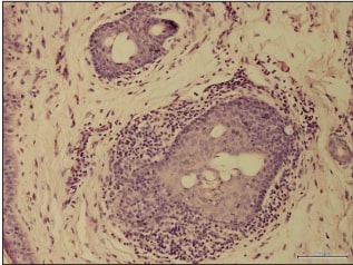
Fig. 1. Active stage. Proliferation of epithelial cells of a deformed hair follicle; perifollicular inflammatory infiltration. Haematoxylin and eosin staining. Magnification: objective 40, eyepiece 10.
In the skin biopsies of patients with the chronic stage of AA, fibroplastic and atrophic processes predominated. The epidermis was also unevenly thickened, with more marked acanthosis, para- and hyperkeratosis and proliferation of the basal layer epithelium. Scanty perifollicular and perivascular infiltrates consisting of lymphocytes, histiocytes and plasma cells were found, along with perifollicular and perivascular sclerosis, sclerosis of the dermis with thickened collagen fibres and fibrous strands, and dermal oedema. The walls of most vessels were thickened, up to obliteration of the vessels. In rare cases signs of angiogenesis were observed. A reduction in the number of HF and their atrophy were noted. In some HF a thin hair shaft was found in which melanin and melanocytes from the hair bulb disappeared (fig. 2).
In the group of patients with the active stage of AA, the numbers of CD4+ T lymphocytes (15.3±0.12 cells) and CD8+ T lymphocytes (25.1±0.26 cells) in the inflammatory infiltrate of the dermis in the fields of view studied were significantly increased compared with the control (p < 0.01). The immunoregulatory index (IRI) was (0.61±0.06), significantly lower than in the control (p < 0.01) (1.08±0.08). CD4+ T lymphocytes were scattered, mainly around follicles and vessels. Occasionally they were found in the dermis under the epidermis. Cytotoxic CD8+ T lymphocytes appeared as compact clusters, also around follicles and vessels, less often in the dermis, and as single cells in the interepithelial zone of the epidermis. Moreover, the more compact expression of CD8+ T lymphocytes was localised in superficial follicles in which hair shafts were absent (fig. 3).
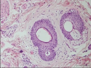
Fig. 2. Chronic stage. Marked perifollicular sclerosis. Haematoxylin and eosin staining. Magnification: objective 40, eyepiece 10.
In the chronic stage of AA, analysis of lymphocyte marker expression showed a significant decrease in the numbers of positive CD4+ and CD8+ T lymphocytes compared with the active stage of AA – (11.7±0.22) (CD4+) and (15.7±0.18) (CD8+) positive cells (p < 0.01). The IRI rose compared with the active stage and was (0.75±0.02). However, the value of this indicator was not significant (p > 0.01).CD56+ expression was found mostly in the inflammatory infiltrate in the active stage of AA. The largest number of CD56+ positive cells in the zone of the inflammatory infiltrate was observed near preserved follicles, and fewer near damaged follicles, averaging (18.00±2.46) positive cells. The intensity of cytoplasmic staining was rated as weak (fig. 4). In the chronic stage of AA, the number of CD56+ positive cells decreased significantly (7.56±0.98) compared with the active stage (p < 0.01), but remained higher than in the control group. A weak reaction was noted in the epithelium and a more intense one in the nerve trunks.
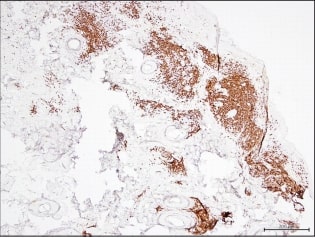
Fig. 3. Skin fragment. Active stage of AA. Expression of CD8+ T lymphocytes located perifollicularly and perivascularly in the dermis. IHC reaction. FLEX visualisation system. Magnification: objective 4, eyepiece 10.
To study in detail the level and quality of the macrophage component of the inflammatory infiltrate in the skin of AA patients, an IHC reaction was performed with the marker of macrophage lineage, CD68+. In the IHC reactions in skin biopsies, macrophages were scattered within the inflammatory infiltrate and lined up in a chain along the basal layer of the follicular epithelium; single macrophages appeared in the interepithelial zone. CD68+ cells showed granular staining of the cytoplasm and varied in size, being mostly large, with marked intense cytoplasmic staining. In the active stage of AA the number of CD68+ macrophages was (29.20±4.06). In the chronic stage of AA the number of macrophages decreased, and the intensity of cytoplasmic staining and its granularity fell. The number of CD68+ macrophages was (16.20±2.34). The values in the active and chronic stages were significantly changed relative to the control and between the groups (p < 0.01).
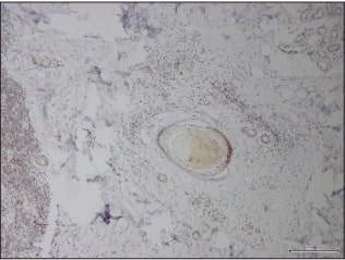
Fig. 4. Skin fragment. Active stage of AA. CD56 expression in cells of the inflammatory infiltrate near a preserved hair follicle. IHC reaction. FLEX visualisation system. Magnification: objective 20, eyepiece 10.
The content of TNF-α was (6.60±0.68) cells in the fields of view studied in the active stage and (4.50±0.38) cells in the chronic stage of AA. These data differ statistically significantly from the control (1.10±0.29) and between the groups (p < 0.01). In analysing expression with the IL-1 marker, cytoplasmic expression in macrophages and lymphocytes was also noted in the active stage of AA. The number of IL-1 cells was (3.70±0.45) in the fields of view studied. IL-1 expression in the chronic stage was (2.10±0.34) cells. Both values differed significantly from the value in the control group (0.70±0.08, p < 0.01) and did not differ significantly from each other (p > 0.01). A similar IHC picture was seen in the analysis of slides with the IL-6 marker, which in the active stage of AA appeared as cytoplasmic staining of macrophages and lymphocytes and was 4.20±0.45 cells, significantly higher (p < 0.01) than in the chronic stage – (2.70±0.36). These values differed significantly (p < 0.01) from the control (1.30±0.08).
AA is characterised by a disturbance of the HF growth cycle: inflammatory cells (CD8+ T cells, CD68+, NK cells and others) attack anagen hair follicles. An increase in the number of inflammatory cells around the HF leads to their miniaturisation and a shortened hair growth cycle with transition to kenogen. The severity of the infiltrate correlated with the acuteness of the pathological process and was more marked in samples from the active stage of AA. In the chronic stage of AA, fibroplastic and, in places, atrophic processes predominated. Besides fibroblasts, macrophages, one of the body's main cytokine-producing cells, also play an important role in fibrogenesis. An uncontrolled immune response associated with hypercytokinaemia may underlie the pathogenesis of early fibrosis [3]. In chronic inflammation in AA patients, the main role is played by immunocompetent cells such as CD68+ with an admixture of single CD8+ lymphocytes. When AA was active, the number of CD8+ cytotoxic T lymphocytes in the patients' skin increased. According to IHC analysis, activated CD8+ T cells can penetrate the HF, while CD4+ T cells are located almost exclusively in the perifollicular area, which can readily disturb hair growth. The CD4/CD8 ratio fell significantly relative to the control, which explains the reduction of local cellular immunity in the lesion. In addition, the increased number of macrophages in the inflammatory infiltrate of the skin indicates an immune response and persistence of chronic inflammation, which may serve as a diagnostic marker not only of the classic manifestations of AA but also in assessing the effectiveness of treatment.
Single NK cells were observed around healthy anagen HF, while follicles in the areas of damage in AA showed visible clusters of CD56+ cells. The marked presence of positive NK cells (CD56+) in the inflammatory infiltrate in the active stage of AA should be noted. Recent studies have revealed overexpression of ULBP3 (cytomegalovirus-binding protein) on chromosome 6 (6q25) in AA, which encodes activation of the ligand of the NK cell receptor NKG2D on natural killer cells and initiates an autoimmune response. ULBP3-encoded proteins are present in large amounts in damaged HF. The proteins recruit NK cells carrying NKG2D receptors. Across different organs there is a common mechanism responsible for the expression of signalling proteins for NKG2D receptors, which triggers the destruction of the cell. These genes are responsible for the development of other autoimmune diseases, such as rheumatoid arthritis and type 1 diabetes. These discoveries are regarded as promising subjects in the study of therapy, including in AA [13, 15].
A high content of CD8+ lymphocytes, through cooperation with T helper cells (CD4+) and macrophages, forms a T cell-mediated cytotoxic immune response associated with NK cells [15]. The inflammatory lymphohistiocytic infiltrate is located not only around the HF but can penetrate into the cells of the hair bulb and surround vessels, which reflects the morphogenesis of autoimmune inflammation. Immune cytolysis of HF cells is carried out not only by T lymphocytes but also by proinflammatory cytokines such as TNF-α, IL-1 and IL-6. TNF-α is a strong inducer of apoptosis and suppresses keratinocyte proliferation. Cytokines can currently be regarded as a mediator link in the formation of the pathophysiological stage of autoimmune reactions in AA, disrupting the mechanisms that maintain the state of immune tolerance of HF structures [3, 5]. The cytokines studied are produced by cells of both the lymphohistiocytic infiltrate and the HF when these are immunologically damaged.
Conclusion
Thus, summarising the literature data and our morphological study, which used IHC to establish the phenotype of the cells of the inflammatory infiltrate and the reaction of proinflammatory cytokines, it can be stated that the changes in the inflammatory focus depend on the stage of the pathological process. In the active stage of the disease there is a massive inflammatory infiltrate with a predominance of cytotoxic T lymphocytes, CD56+ and CD68+ cells, and an increased content of proinflammatory cytokines (IL-1, IL-6, TNF-α). By contrast, in the chronic stage of AA we observe a minimal degree of cellular response, with a reduced inflammatory infiltrate and a redistribution of the infiltrate cells (CD8+ and CD4+) in favour of fibroblastic and histiocytic elements. The content of the proinflammatory Th-1 cytokines in the chronic stage of AA also falls significantly. These changes reflect a shift of the inflammatory reaction in the AA focus towards reparative and sclerotic mechanisms. The lack of a unified view of the causes and course of AA makes it important to study the links in the pathogenesis of this dermatosis, because the data obtained make it possible to develop pathogenetic approaches to therapy that take the stage of the pathological process into account.
1. Насонова Е.Л. Новые направления фармакотерапии ревматических заболеваний – ингибиция интерлейкина 6 и интерлейкина 17 [Текст] / Е.Л. Насонова // Современная ревматология. – 2013. – No 3. – С. 5-14.
2. Сербина И.М. Цитокинопосредованные механизмы формирования гнездной алопеции [Текст] / И.М. Сербина // Научные ведомости БелГУ. Серия: Медицина. Фармация. – 2014. – Т. 28, No 24 (195). – С. 32-38.
3. Соотношение процессов апоптоза, пролиферации, неоангиогенеза и клеточной дифференцировки при иммунном воспалении в очагах гнездной алопеции [Текст] / А.Г. Гаджигороева, Е.А. Коган, Н.Н. Потекаев, Г.П. Терещенко // Клиническая дерматология и венерология. – 2010. – No 2. – С. 23-31.
4. Сучасні методи діагностики нерубцевих алопецій: Метод. рекомендації [Текст] / Л.А. Болотна, І.М. Сербіна, Ю.С. Овчаренко, Ю.В. Качук. – Харків, 2013. – 24 с.
5. Alopecia areata update: part I. Clinical picture, histopathology, and pathogenesis [Text] / A. Alkhalifah, E. Wang, J. Shapiro [et al.] // J. Am. Acad. Dermatol. – 2010. – Vol. 62. – P. 191-202.
6. Clinico etiological study of Alopecia areata [Text] / G. Bharathi, P. Venkata Ramana, K. Sridevi, G. Usha // Journal of Dental and Medical Sciences. – 2015. – Vol. 14, No.6. – P. 29-32.
7. D’Ovidio R. Alopecia Areata: news on diagnosis, pathogenesis and treatment [Text] / R. D’Ovidio // G. Ital. Dermatol. Venereol. – 2014. – Vol. 149. – P. 25-45.
8. Gilhar A. Collapse of Immune Privilege in Alopecia Areata: Coincidental or Substantial? [Text] / A. Gilhar // Journal of Investigative Dermatology. – 2010. – Vol. 130. – P. 2535–2537.
9. Gilhar A. Alopecia areata [Text] / A. Gilhar, A. Etzioni, R. Paus // The New England Journal of Medicine. – 2012. – Vol. 366. – P. 1515-1525.
10. Hordinsky M. Overview of Alopecia Areata [Text] / M. Hordinsky //Journal of Investigative Dermatology Symposium Proceedings. – 2013. – Vol. 16, No. 1. – P. S13-S15.
11. Investigation of the inflammatory mechanisms in alopecia areata [Text] / D.C. Emel, S. Ekin, U. Meltem [et al.] // Am. J. Dermatopathol. – 2009. – Vol. 31. – P. 53-60.
12. Ito T. Recent Advances in the Pathogenesis of Autoimmune Hair Loss Disease Alopecia Areata [Text] / T. Ito // Clinical and Developmental Immunology. – 2013. – ID348546. – P. 1-6.
13. Petukhova L. Genomewide association study in alopecia areata implicates both innate and adaptive immunity [Text] / L. Petukhova // Nature. – 2010. – Vol. 466. – P. 113-117.
14. Value of transverse section scalp biopsy in alopecia areata - a clinicopathological correlation [Text] / K. Jameel, A. Ejaz, M. Sohail, S.B. Rahman // J. Col. Phys. Surg. Pak. – 2008. – Vol. 18, No. 6. – P. 338-341.
15. Wang E. Etiopathogenesis of alopecia areata: Why do our patients get it? [Text] / E. Wang, J. McElwee // Dermatologic Therapy. – 2011. – Vol. 24. – P. 337-347.
References
2. Serbina, I.M. (2014) Tsitokinoposredo- vannyie mehanizmyi formirovaniya gnezdnoy alopetsii. Nauchnyie vedomosti BelGU. Seriya: Meditsina. Farmatsiya. 28, (24 (195)), 32-38. (in Russian)
3. Gadzhigoroeva, A.G., Kogan, E.A., Pote- kaev, N.N., Tereschenko, G.P. (2010) Soot- noshenie protsessov apoptoza, proliferatsii, neoangiogeneza i kletochnoy differentsirovki pri immunnom vospalenii v ochagah gnezdnoy alopetsii. Klinicheskaya dermatologiya i ven- erologiya. 2, 23-31. (in Russian)
4. Bolotna,L.A., Serbina,I.M., Ovcharenko, Yu.S., Kachuk, Yu.V. (2013) Suchasni metody diahnostyky nerubtsevykh alopetsii: Metod. rekomendatsii. 24. (in Ukrainian)
5. Alkhalifah, A., Wang, E., Shapiro, J. [et al.] (2010) Alopecia areata update: part I. Clini- cal picture, histopathology, and pathogenesis. J. Am. Acad. Dermatol. 62, 191-202.
6. Bharathi, G., Venkata Ramana, P., Sridevi, K., Usha, G. (2015) Clinico etiologi- cal study of Alopecia areata. Journal of Dental and Medical Sciences. 14 (6), 29-32.
7. D’Ovidio, R. (2014) Alopecia Areata: news on diagnosis, pathogenesis and treatment. G. Ital. Dermatol. Venereol. 149, 25-45. 8. Gilhar, A. (2010) Collapse of Immune Privilege in Alopecia Areata: Coincidental or Substantial? Journal of Investigative Dermatol- ogy. 130, 2535-2537.
9. Gilhar, A., Etzioni, A., Paus, R. (2012) Alopecia areata. The New England Journal of Medicine. 366, 1515-1525.
10. Hordinsky, M. (2013) Overview of Alo- peciaAreata. Journal of Investigative Dermatol- ogy Symposium Proceedings. 16 (1), S13-S15.
11. Emel, D.C., Ekin, S., Meltem, U. [et al.] (2009) Investigation of the inflammatory mechanisms in alopecia areata. Am. J. Dermato- pathol. 31, 53-60.
12. Ito, T. (2013) Recent Advances in the Pathogenesis of Autoimmune Hair Loss Disease Alopecia Areata. Clinical and Developmental Immunology. ID348546, 1-6.
13. Petukhova, L. (2010) Genome-wide association study in alopecia areata implicates both innate and adaptive immunity. Nature. 466, 113-117.
14. Jameel, K., Ejaz, A., Sohail, M., Rah- man, S.B. (2008) Value of transverse section scalp biopsy in alopecia areata - a clinicopatho- logical correlation. J. Col. Phys. Surg. Pak. 18 (6), 338-341.
15. Wang, E., McElwee,J. (2011) Etiopatho- genesis of alopecia areata: Why do our patients get it? Dermatologic Therapy. 24, 337-347.