
Dermatosurgery

Dermato-oncologist is a doctor who specializes in the diagnosis, treatment, and prevention of skin growths, including malignant ones.
Most malignant skin tumors show no pain in the early stages and can look like an ordinary mole.
That is why early diagnosis is one of the key factors for successful treatment: a timely visit allows abnormal changes to be detected at an early stage and significantly increases the chance of full recovery.
Consultations are conducted in a modern clinic using high-tech diagnostic methods.
Dermato-oncologist appointments in Kharkiv are held at the Institute of Trichology clinic, located at 17 Danilevsky Street.
WHAT WE DIAGNOSE AND TREAT
- Melanoma
- Basal cell carcinoma (basalioma)
- Squamous cell carcinoma
- Actinic keratosis
- Melanocytic nevus (mole)
- Dermatofibroma
- Juvenile xanthogranuloma
- Seborrheic keratosis
- Angioma
- Papillomas
Diagnostics and procedures
Make an appointment
DERMATO-ONCOLOGIST CONSULTATION
The consultation includes a comprehensive examination of the entire skin surface to identify both malignant and benign growths with a risk of malignant transformation.
During the appointment, the doctor:
- examines the entire skin surface
- performs dermoscopy
- assesses risks
- forms a plan of action (observation / removal / biopsy)
WHEN A DERMATO-ONCOLOGIST CONSULTATION IS NEEDED
You should see a doctor if any suspicious changes appear on the skin or mucous membranes. Early diagnosis helps prevent disease progression, reduces the risk of complications, and simplifies treatment.
A consultation is recommended if:
- a growth appears that differs from others;
- a change in shape, size, or color is observed;
- itching, burning, or discomfort occurs;
- bleeding or other discharge appears;
- redness or flaking occurs;
- soreness appears.
RISK FACTORS
A preventive examination is recommended even without complaints, especially with the following risk factors:
- more than 50 nevi;
- fair skin phototype (I-II);
- past episodes of sunburn;
- hereditary predisposition to skin cancer;
- age over 50;
- immunosuppressive conditions;
- regular use of a tanning bed.
PREPARING FOR THE APPOINTMENT
A few days before the consultation:
- do not apply medicated ointments;
- avoid products that change skin color.
Before the visit:
- do not apply cosmetic products;
- avoid tanning and skin injury.
On the day of the appointment:
- skin should be clean;
- bring any previous medical reports and test results (if available).
DIAGNOSTIC METHODS
The following methods are used for accurate skin assessment:
- Digital dermoscopy - detailed analysis of growth structure using magnification and digital imaging.
- Punch biopsy - tissue sampling for further histological examination.
- Mole mapping - full-body skin photography with subsequent digital analysis and dynamic monitoring option.
- Laboratory tests - used to clarify the diagnosis and monitor treatment.
In complex clinical cases, a multidisciplinary consultation is held involving international experts.
TREATMENT METHODS
Treatment strategy is determined individually based on the clinical situation and the patient's characteristics:
- Radiowave removal
- Shave excision
- Cryotherapy
- Laser removal
- Surgical treatment
- MOHS surgery
- Topical therapy
- PUVA therapy
- Systemic treatment
- Biological therapy

WHAT THE PATIENT RECEIVES
Accurate diagnosis
- Comprehensive skin assessment using modern imaging methods, namely digital dermoscopy.
Modern treatment approaches
- Application of evidence-based medicine, modern clinical protocols, and up-to-date treatment methods.
Comfort and safety
- Organization of the treatment process taking into account the patient's individual needs and adherence to high safety standards.
Doctors
Price
Dermato-oncologist consultation | |
| Consultation of a dermato-oncologist with digital dermatoscopy using FotoFinder equipment | from 1500 UAH |
| Pre-operative consultation with a dermato-oncologist | 1200 UAH |
| Follow-up consultation with a dermato-oncologist | 800 UAH |
| Dermato-oncologist consultation during a course of topical immunotherapy (TIT) | 800 UAH |
| Digital mole mapping on FotoFinder equipment | 3000 UAH |
Dermato-oncology diagnostics | |
| Punch biopsy of the trunk / limbs, per lesion | 1700 UAH |
| Punch biopsy of the trunk / limbs, from the 2nd lesion | 1300 UAH |
| Punch biopsy in anatomically complex areas, per lesion | 2500 UAH |
| Punch biopsy in anatomically complex areas, each further lesion from the 2nd | 1700 UAH |
| Excisional biopsy of a skin tumor with assessment of resection margins | from 3500 UAH |
| Histopathological examination of the skin in benign, malignant and tumor-like processes | from 1500 UAH |
Minimally invasive dermatosurgical removal | |
| Shave removal of skin lesions in anatomically complex areas, per lesion | 1200 UAH |
| Shave removal of skin lesions in anatomically complex areas, from the 2nd lesion | 800 UAH |
| Shave removal of skin lesions on the trunk / limbs, per lesion | 800 UAH |
| Shave removal of skin lesions on the trunk / limbs, each further lesion from the 2nd | 500 UAH |
| Radio-wave removal of benign skin lesions (keratomas / angiomas) on the face, 1 zone | 3000 UAH |
| Radio-wave removal of benign skin lesions (keratomas / angiomas) on the trunk / limbs (1 zone) | 2500 UAH |
| Radio-wave papilloma removal, per lesion | 350 UAH |
| Radio-wave papilloma removal on the eyelids, per lesion | 500 UAH |
| Radio-wave papilloma removal on the eyelids (package) | 3000 UAH |
| Radio-wave papilloma removal, 1 zone (package) | 2000 UAH |
| Radio-wave removal of genital warts in anatomically complex areas, per lesion | 600 UAH |
| Radio-wave removal of genital warts in anatomically complex areas (1 zone) | 3500 UAH |
| Radio-wave removal of seborrheic keratomas / angiomas, per lesion | 600 UAH |
| Radio-wave removal of seborrheic keratomas / angiomas, each further lesion from the 2nd | 400 UAH |
| Radio-wave removal of seborrheic keratomas / angiomas (1 zone) | 3000 UAH |
Surgical removal of skin lesions | |
| Surgical removal of skin lesions, complexity level I | 3500 UAH |
| Surgical removal of skin lesions, complexity level II | 5000 UAH |
| Surgical removal of skin lesions, complexity level III | 7000 UAH |
| Surgical removal of skin lesions, complexity level I, in anatomically complex areas | 5000 UAH |
| Surgical removal of skin lesions, complexity level II, in anatomically complex areas | 7000 UAH |
| Surgical removal of skin lesions, complexity level III, in anatomically complex areas | 9000 UAH |
| Oncological reconstruction of the post-operative wound, complexity level I | 9000 UAH |
| Oncological reconstruction of the post-operative wound, complexity level II | 10000 UAH |
| Oncological reconstruction of the post-operative wound, complexity level III | 11000 UAH |
| Oncological reconstruction of the post-operative wound, complexity level I, in anatomically complex areas | 13000 UAH |
| Oncological reconstruction of the post-operative wound, complexity level II, in anatomically complex areas | 15000 UAH |
| Oncological reconstruction of the post-operative wound, complexity level III, in anatomically complex areas | 18000 UAH |
Re-excision surgery (with local tissue repair) | |
| Linear closure with resection margins extended up to 5 mm | 11000 UAH |
| Linear closure with resection margins extended up to 1 cm | 13000 UAH |
| Linear closure with resection margins extended up to 2 cm | 15000 UAH |
| Flap repair with resection margins extended up to 5 mm | 13000 UAH |
| Flap repair with resection margins extended up to 1 cm | 15000 UAH |
| Flap repair with resection margins extended up to 2 cm | 18000 UAH |
FAQ
A dermato-oncologist is a doctor who specialises in diagnosing, treating and preventing skin growths, including malignant ones: melanoma, basal cell carcinoma and squamous cell carcinoma. They also assess benign growths that need monitoring or removal: moles, seborrheic keratoses, dermatofibromas, angiomas and papillomas.
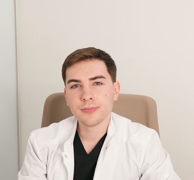
A dermatologist treats skin diseases in general: dermatitis, psoriasis, acne, fungal infections. If you are worried about a mole or a new growth, a dermato-oncologist is the right specialist. At the clinic you will see Nazar Tsalin, MD, a dermatovenereologist, dermato-oncologist and surgical oncologist, member of the International Dermoscopy Society and the EADV.
Book a check if you notice:
- a new growth that looks different from the others;
- a change in the size, shape or colour of a mole, uneven borders, new shades;
- bleeding, a crust that keeps coming back, or a sore that does not heal;
- itching, burning, tenderness, redness or flaking;
- a new dark streak under a nail with no obvious cause.
No pain does not mean the growth is safe: malignant skin tumours often do not hurt in the early stages and can look like an ordinary mole. No single sign confirms cancer, but each is a reason for an examination. More in What Skin Cancer Looks Like: Early Signs.
Yes, a preventive check is recommended even without complaints, especially if you have more than 50 moles, fair skin (phototype I–II), past sunburns, a family history of skin cancer, are over 50, have a weakened immune system or use tanning beds regularly.
There is no single interval for everyone. For a healthy person with no relevant history, a check once a year is the usual recommendation. People with many moles, atypical naevi or a past melanoma may be advised regular monitoring with dermoscopy or mole mapping. If a mole starts to change, do not wait for the planned visit. More in Mole and Skin Lesion Checks in Kharkiv.
The doctor asks when the growths appeared and how they changed, and whether you have had skin cancer, sunburns or melanoma in the family. Then the doctor examines the whole skin, not only the mole that worries you, and performs digital dermoscopy of the growths that need assessment.
Dermoscopy is painless and does not damage the skin. Based on the results, the doctor assesses the risks and proposes a plan: monitoring, a follow-up check comparing images, a biopsy, or removal with histological examination. Patients at higher risk may be advised digital mole mapping. See current prices for the consultation and diagnostics in the price list.
No complex preparation is needed, but a few rules help the doctor assess your skin accurately:
- for a few days before, do not apply medicated ointments or products that change skin colour to the growths;
- before the visit, do not use make-up or tinted products, and avoid tanning and skin injury;
- on the day of the appointment your skin should be clean.
Bring previous medical reports, test results, histology reports and photos of the growths, if you have them. Tell the doctor whether the mole has changed, bled, itched or been injured.
A full skin check is a standard part of a dermato-oncologist consultation. Suspicious changes are sometimes not where the patient expects them: a person comes in about a benign growth that gets in the way or keeps being injured, and during the check the doctor notices another one they did not know about.
It also matters to compare moles with each other: a growth that clearly looks different from the rest deserves special attention. So if you have never had a check, the doctor may suggest looking at other areas too, especially those that are hard to see yourself: the back, the scalp, the feet.
They are simple guides for checking pigmented spots yourself. The ABCDE rule: A for asymmetry, B for uneven or blurred borders, C for uneven colour, D for a diameter over 6 mm, E for evolution, meaning any change over time. Change matters most: a melanoma can be smaller than 6 mm.
The ugly duckling sign means that a mole which looks clearly different from the others on your body may be suspicious. Both rules help you see a doctor in time, but they cannot rule out cancer on their own and are not meant for judging basal cell or squamous cell carcinoma. More in What Skin Cancer Looks Like: Early Signs.
Yes. Size is only one criterion: in the early stage a melanoma can be small, and some types do not have the typical dark colour. The risk of many skin cancers grows with age, but melanoma also occurs in young people.
So it is more important to watch for change than for size: asymmetry, borders, colour, structure, bleeding. If a growth is changing, have it checked sooner rather than waiting until it gets bigger or starts to hurt. Read about common patient misconceptions in Myths About Moles and Skin Cancer.
Basal cell carcinoma (basalioma) is one of the most common skin cancers. It most often appears on the face, nose, ears, neck and scalp. It can look like a pearly or pink bump with tiny blood vessels, a pink scaly patch, a spot that will not go away, or a sore that crusts over again and again.
It grows slowly and very rarely spreads to distant organs, but without treatment it gradually destroys the surrounding tissue, so a larger operation may be needed later. The diagnosis is confirmed by dermoscopy and histological examination. More in Basal Cell Carcinoma Removal in Kharkiv.
Both mostly appear on skin that has had years of sun: the face, ears, lips, hands and a thinning scalp. Actinic keratosis usually looks like a rough, dry patch and is considered a precancerous change, so it is treated and monitored. Squamous cell carcinoma can show up as a scaly patch that does not go away, a firm bump, a crusted growth or a sore that does not heal; in some cases it can spread to other tissues.
They cannot be told apart by eye, so do not treat such a patch with ointments or burn it off yourself. The doctor will assess it with dermoscopy and order a biopsy if needed. Treatment options for actinic keratosis include photodynamic therapy.
Do not panic: an accidental injury does not mean melanoma will develop. Clean the wound with an antiseptic, do not pick the crust and do not try to burn the growth off.
That said, an injured mole is one of the reasons for dermoscopy. The doctor will see how it is healing and check it for suspicious features. If a mole is constantly rubbed by clothing, a belt, a razor or a bra strap, you can discuss planned removal after the check. Do not delay the visit if the injured growth keeps bleeding, does not heal or changes in appearance.
No. Modern medicine does not support the idea that a properly performed biopsy or surgical removal of a suspicious growth causes cancer to spread. On the contrary, a diagnostic procedure is an important step towards a diagnosis and the right treatment.
The real danger lies elsewhere: a malignant growth is removed without prior assessment, for example in a beauty salon with a laser or another method that destroys the tissue. Then nothing is left for histology and the diagnosis comes late. So fear of removal should not be a reason to put off an examination. More in Myths About Moles and Skin Cancer.
Cutting it off with a blade or scissors, tying it with thread, or burning it with celandine, acids, alkalis or wart removers can cause bleeding, infection, chemical burns, incomplete removal and scarring. Most importantly, it makes a diagnosis impossible. If the growth turns out to be malignant, self-treatment delays the right diagnosis.
Likewise, ointments cannot rule out melanoma: less inflammation or a changed surface does not mean the problem is gone. If a growth is changing, bleeding or not healing, show it to a doctor instead of trying to get rid of it at home.
Mole mapping (digital skin mapping, Total Body Mapping) means photographing the entire body surface and recording individual growths with dermoscopy. The aim is not just to take pictures but to compare the skin over time and spot new or changed moles.
It is most useful for people with many moles, atypical naevi, a past melanoma or melanoma in the family. If you have few moles and no risk factors, a regular check with dermoscopy is often enough. Whether you need mapping is the doctor's call. More in Mole Mapping in Kharkiv: Digital Skin Mapping.
The examination has several steps: the doctor reviews your history and risk factors, takes a series of standardised photos of different body areas, examines individual growths with a digital dermatoscope and interprets the results. Depending on the number of growths, it usually takes about 30–45 minutes. It is non-invasive and painless.
The images are stored in a secure database and compared with new ones at your next visit. After the dermoscopic examination you receive a printout of the results with images and recommendations. Keep these documents and bring them to later consultations, including if you plan to be examined at another medical facility.
A photo can be a reason to book a check, but it does not replace one. Image quality depends on lighting, focus and angle, and an ordinary camera does not give dermoscopic magnification. Apps and AI algorithms can be wrong both ways: calling a malignant growth benign, or the other way round.
Even in professional digital dermoscopy systems, the software only helps the doctor organise images. The diagnosis is made by a specialist who considers your history, changes over time, the clinical picture and, when needed, histology. Photographing your moles to track them is useful, but if something changes, come in for dermoscopy.
No. A biopsy is ordered precisely when an examination and dermoscopy are not enough to say for sure what a growth is. The result may confirm a benign growth, show precancerous changes or reveal a malignant process, and the next steps depend on it.
A punch biopsy is often used: under local anaesthesia a small cylinder of tissue is taken with a special instrument, and a dressing is applied. Sometimes the doctor recommends complete diagnostic excision of the growth straight away. The tissue is examined by histology laboratories in Ukraine and Germany that the clinic works with. More about skin biopsy.
Not always, but for suspicious growths it is decisive. Histology means examining the tissue under a microscope. It is what confirms or rules out melanoma or another skin cancer and, if the process is malignant, defines the tumour features that guide further treatment.
After removal of a typical benign growth, the doctor decides whether histology is needed based on the clinical picture, dermoscopy and the method used. But if malignancy is suspected or the growth has atypical features, the tissue must go to a pathologist. Do not consider the matter closed just because the growth is gone from your skin: it is important to know what exactly was removed.
The department uses radiowave removal, shave excision, cryotherapy, laser removal, surgical excision and MOHS surgery. There is no universal method: the choice depends on the type of growth, its size, location, dermoscopic picture and whether histology is needed.
The doctor first assesses the growth, then chooses the method. If melanoma is suspected, the priority is complete removal with tissue kept for examination. Laser, cryodestruction and other methods that destroy tissue are not suitable then, because nothing is left for histology. That is why a suspicious mole should not be removed in a beauty salon without diagnosis. More in Mole Removal in Kharkiv.
The approach depends on the tumour's size and location, histological type, how clear its borders are, previous treatment and the state of the immune system. For most basal cell carcinomas the main method is complete surgical removal with histological examination. In selected low-risk cases topical therapy, cryosurgery or other methods may be considered, but they do not suit everyone.
MOHS surgery removes the tumour layer by layer, checking the margins of each layer under a microscope until they are clear. It is especially valuable on the nose, eyelids and lips, where healthy tissue must be spared as much as possible. No method can guarantee that the tumour will never return, so follow-up checks are needed after treatment. More in Basal Cell Carcinoma Removal in Kharkiv.
Diagnostic excision and further treatment are separate stages. Once the diagnosis is confirmed, the doctor reviews the pathology report: the tumour type, Breslow thickness, whether there is ulceration and other features. These determine whether a repeat, wider excision is needed, what extent of surgery is indicated and whether further tests are required.
The plan follows current clinical guidelines. In complex cases a multidisciplinary consultation with international experts is held. It is important not to delay the next steps and to attend the follow-up checks your doctor schedules.
Not necessarily. In children, moles can appear and gradually grow in proportion to the body, and this alone is not a sign of disease. Melanoma in childhood is extremely rare, and changes in children's moles are assessed in the context of age and the overall clinical picture.
Do bring your child to a doctor if a mole changes quickly, gets injured, bleeds or looks clearly different from the others. Children with a congenital mole are advised to have dermoscopy once a year to track it over time. The check and dermoscopy are painless, so children cope with them easily.
Yes. Dermoscopy is safe during pregnancy and breastfeeding: it is an examination with an optical system and polarised light that does not damage the skin. Some researchers note that moles may become more active during pregnancy or after childbirth, so a screening check during this time makes sense.
Always tell the doctor that you are pregnant or breastfeeding: the next steps depend on it. Some planned procedures, such as radiowave removal of benign growths, are not done during this period. If a growth is suspicious, the doctor decides on the approach individually without delaying the necessary diagnosis.
Dermato-oncologist consultations take place in person at the Institute of Trichology clinic in Kharkiv, 17 Danilevsky Street: assessing growths requires a skin examination and dermoscopy.
To get the most from the visit, bring all previous reports, test results, dermoscopy images or mapping reports, histology reports and, if possible, photos showing how the growths have changed. This helps the doctor compare the new findings with earlier ones. In complex cases a multidisciplinary consultation with international experts is held. About the doctor: Nazar Tsalin, MD, dermato-oncologist and surgical oncologist.
The main risk factor for basal cell carcinoma and other skin cancers is ultraviolet radiation: over a lifetime it builds up DNA damage in skin cells. Frequent sunburns, long unprotected time in the sun and tanning beds also raise the risk.
What helps: broad-spectrum sunscreen, clothing and hats, shade, and giving up tanning beds. Sun-protection habits are best built from childhood. People who work outdoors a lot, such as military personnel and construction workers, need to be especially careful. And even with good protection, do not skip regular skin checks.
