
Digital Mole Mapping
Digital mole mapping is an advanced way to document skin growths and track them over time, using the FotoFinder professional system.
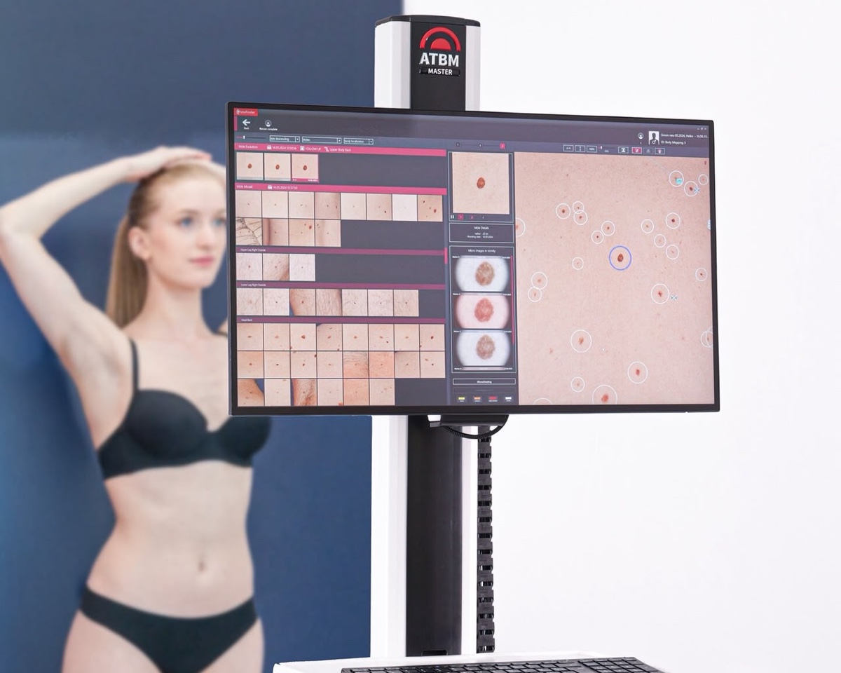
During the procedure we take standardized photographs of the body surface that record where your many skin growths are and how they look. When needed, individual moles are also documented with digital dermoscopy.
The stored images are used for comparison at later visits: the doctor can see whether new growths have appeared and whether existing moles have changed in size, shape, color or dermoscopic structure.
Who mole mapping is recommended for
The method is especially recommended for patients at higher risk of melanoma, including:
- patients with many moles (more than 50);
- people with a genetic predisposition to melanoma;
- patients with multiple dysplastic nevi and a personal history of melanoma;
- patients with other risk factors defined by international guidelines.
Make an appointment
The goal of mapping
The main goal of mapping is systematic monitoring of your skin: spotting new or changed growths in time and improving the chances of early diagnosis of melanoma and other skin cancers.
For patients with no indications for full digital mapping, a regular examination by a dermato-oncologist with digital dermoscopy of the individual growths that need a closer look may be enough.
Doctors
Prices
| Digital mole mapping on FotoFinder equipment | 3000 UAH |
Frequently Asked Questions
Mapping uses standardized photographs of the body surface, so the location and look of all your growths are recorded at once. Individual moles can also be imaged with a dermatoscope. At the next visit the doctor compares the new images with the stored ones.
Whether new growths have appeared and whether existing moles have changed in size, shape, color or dermoscopic structure.
Patients at higher risk of melanoma: more than 50 moles, a genetic predisposition to melanoma, multiple dysplastic nevi and a personal history of melanoma, or other risk factors from international guidelines.
Then a regular examination by a dermato-oncologist with digital dermoscopy of the growths that need a closer look may be enough.
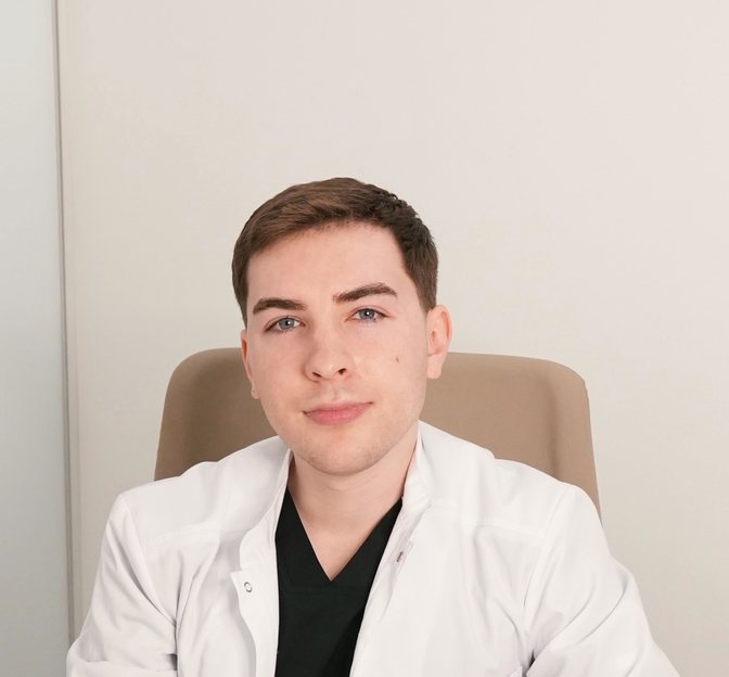
Dr. Nazar Tsalin explains the method in his article “Mole Mapping”.
