Trichoscopic differential diagnostic algorithms for non-scarring alopecias
co-authored with Inessa Serbina and Bohdan Lytvynenko
Introduction
Dermoscopy is now firmly established in the diagnosis of diseases of the hair and scalp as a non-invasive and effective method [19]. The term "trichoscopy" was introduced in 2006 by the American researcher L. Rudnicka. Knowledge of dermoscopic signs opens up new possibilities in the study and diagnosis of hair diseases. Examining the scalp with a dermatoscope provides valuable clinical information about the actual state of the skin. The method can be used both as an auxiliary tool in the differential diagnosis of hair loss and scalp diseases and at the follow-up stage during treatment[2, 3,18,19].
Trichoscopy requires special equipment: an integrated diagnostic system that combines diagnostic computer software and video diagnostic hardware, a videodermatoscope or trichoscope with a working magnification from x10 to x1000 [4, 19]. UV-enhanced (UV-reflected) trichoscopy is a method of examining the hair and scalp first described by a group of authors in 2011 [20]. It differs from classic trichoscopy in that uniform or polarised light is replaced by UV rays with a wavelength covering the spectrum of the Wood's lamp. This method can extend the diagnostic capabilities of trichoscopy in microsporia and pityriasis versicolor [23].
Non-scarring hair loss, which includes androgenetic alopecia, diffuse alopecia, alopecia areata and trichotillomania, accounts for more than 80% of all hair diseases, making it the most common cause of hair loss; it affects from 30–40% of people under the age of 50 [1, 4]. Diagnostic trichoscopic criteria for non-scarring alopecias have now been identified; knowing them reduces the number of diagnostic errors and makes it possible to prescribe appropriate and timely treatment in each case[11, 18].
A trichoscopic diagnosis is based on assessing the hair shaft, the follicular openings and the perifollicular epidermis.
Assessing the hair shaft
A classification of hair shaft abnormalities seen on trichoscopy has been proposed, which distinguishes the following forms: fractured shafts, narrowings, hairs with nodular structures, coils and twists, bands, and short hairs [19, 20]. Many types of short hair are decisive in the differential diagnosis of the types of baldness most common in clinical practice. Such hairs are less than 5 mm long. They include hypopigmented vellus hairs, which are most typical of androgenetic alopecia 3,4. Vellus hairs can also be seen in alopecia areata, especially during regression of the disease [7, 25]. Another type of short hair is comma hairs and corkscrew hairs, which are typical of scalp microsporia. Short flame hairs are found in trichotillomania [19, 20]. Exclamation mark hairs are narrowed at the proximal end. This type of abnormality is seen in alopecia areata and, less often, in trichotillomania [9].
Assessing the follicular openings (dots)
Trichoscopy makes it possible to distinguish normal, empty and fibrotic follicles, or follicles containing biological material such as hyperkeratotic plugs or hair residues. Dots is a general term for the openings of hair follicles seen on trichoscopy [18].
Black dots (formerly called cadaverised hairs) are pigmented hairs broken or destroyed at scalp level. They are seen in alopecia areata, dissecting cellulitis, scalp microsporia and trichotillomania [10, 18, 20].
Yellow dots are hair follicle openings that contain keratin and sebaceous material [12,18]. They are typical of alopecia areata, discoid lupus erythematosus and androgenetic alopecia [9,16]. Rarely, yellow dots can be seen in telogen hair loss and trichotillomania [19].
There are 2 types of white dots: classic ones, which are large and irregular in shape, and small ones. Classic white dots are areas of perifollicular fibrosis and are most often seen in lichen planus [18,19]. Small white dots, which correspond to the openings of hair follicles and exocrine glands, are seen against a pigmented background. They are found in patients with darker skin phototypes, whether or not they have hair loss [6].
Red dots can be seen in discoid lupus erythematosus and are considered a good prognostic sign, pointing to possible hair regrowth [24]. Symmetrically arranged grey or brownish-grey dots are a characteristic sign in the eyebrow area of patients with frontal fibrosing alopecia [19].
Assessment of the perifollicular epidermis covers abnormalities in the colour and structure of the scalp skin that can be seen on trichoscopy, including scaling, colour changes, changes in the surface structure of the skin, and secretion [21].
Assessing the blood vessels
On trichoscopy, the dermal microvessels can vary in type, arrangement and number depending on the disease. Analysing the arrangement of the vessels is particularly important in the differential diagnosis of inflammatory scalp diseases such as psoriasis, seborrhoeic dermatitis and discoid lupus erythematosus [17,19].
Differential diagnosis of non-scarring alopecias
Telogen hair loss (telogen effluvium)
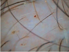
The term telogen hair loss (telogen effluvium) covers a wide range of clinical conditions that share one feature: sudden, extensive shedding of telogen hairs [1,4]. The condition is considered the most common type of hair loss, yet evidence-based knowledge about it remains limited. Trichoscopic signs of telogen effluvium include empty hair follicles, a predominance of single-hair follicular units, perifollicular discolouration (the peripilar sign) and regrowing hairs (Fig. 1). Trichoscopic findings do not depend on the factors that triggered the telogen hair loss[10].
Androgenetic alopecia
Male-pattern and female-pattern androgenetic alopecia share similar trichoscopic signs. Variation in hair shaft thickness, with thin, medium and thick hairs present at the same time, is the most characteristic feature of androgenetic alopecia (Fig. 2). Hair diameter diversity has been shown to reflect follicle miniaturisation in androgenetic alopecia [8,13]. For diagnosis and differential diagnosis in clinical practice there is no need to measure hair shaft thickness precisely, but it can be useful for monitoring treatment efficacy, and it is a prerequisite for clinical trials [14].
Another sign of androgenetic alopecia is an increased proportion of vellus hairs. In the frontoparietal area of patients with female-pattern androgenetic alopecia, vellus hairs make up 20.9% ± 12% [16]. This is considerably higher than the 6.15% ± 4.6% found in healthy individuals [15,16]. Multiple vellus hairs can also be present in alopecia areata, especially during regression of the disease [9,10]. The presence of vellus hairs alone should therefore not be taken as androgenetic alopecia.
In androgenetic alopecia the number of hairs per follicular unit is reduced. In these patients, especially at a late stage of the disease, follicular units with only 1 hair predominate. In the frontoparietal area of patients with female-pattern androgenetic alopecia, such follicular units average 65.2% ± 19.9%. The corresponding figure in healthy individuals is 27.3% ± 13%[25].
Hyperpigmented perifollicular spots (the peripilar sign) are seen in 20% - 66% of patients with androgenetic alopecia. They affect 32.4% ± 4.7% of hair follicle openings [7, 8, 16]. This sign can also be seen in some patients with telogen effluvium and in healthy individuals, the difference being that in androgenetic alopecia the proportion of affected hair follicle openings is higher in the frontal area than in the occipital area. In healthy people and in patients with telogen effluvium the proportion of affected openings is much smaller, and they are spread evenly across all areas of the scalp [15].
The trichoscopy of involutional alopecia matches the signs of androgenetic alopecia: a predominance of single-hair follicular units, reduced hair shaft density with a honeycomb pigment pattern, and a slight tendency to form brown perifollicular spots (the peripilar sign) [19].
Diagnosis and treatment at the Institute
Get a consultation
Alopecia areata
In most cases the diagnosis of alopecia areata is based on clinical signs. However, trichoscopy can help in the differential diagnosis of difficult cases, in particular in the diffuse form of alopecia areata or in patients with complete hair loss [2, 5, 22].
The distinguishing trichoscopic features of alopecia areata are evenly distributed yellow dots, exclamation mark hairs, black dots and regrowing vellus hairs (Fig 3, 4) [2, 4, 19]. The trichoscopic picture of alopecia areata can vary with the activity, severity and duration of the disease. In recent years these differences have been assessed in numerous studies.
Lacarrubba and co-authors examined 200 patients with alopecia areata, divided into the acute and chronic stages of the disease [12]. The study identified 3 signs of acute AA: exclamation mark hairs, black dots and vellus hairs. Inui and co-authors, in a trichoscopic study of 300 patients with AA, found similar markers of disease activity (black dots, tapering hairs and broken hairs) [9]. This study established that vellus hairs are a sign of long-standing inactive disease. Ross and co-authors divided 58 patients with alopecia areata into the following subgroups: patchy, band-like (ophiasis), diffuse and totalis/universalis. The trichoscopic features were similarly expressed in all the subgroups studied and did not depend on the form of alopecia areata [18].
Some patients with long-standing and active alopecia areata have hairs with the typical Pohl-Pinkus constrictions characteristic of monilethrix. In a separate study this type of abnormality was seen in 2 of 66 (3%) patients with alopecia areata examined. Hairs with Pohl-Pinkus constrictions can lead to a misdiagnosis of monilethrix [24].
Signs of hair regrowth in alopecia patches include pigmented short upright hairs and symmetrically coiled hairs [20]. In their publication the authors used the term "pigtail hairs" for hairs that form symmetrical rounded or oval structures as they grow. They should be distinguished from the asymmetrically twisted hairs of trichotillomania.
Trichotillomania
Trichoscopy shows reduced hair density, hairs broken at different lengths, short hairs with trichoptilosis (split ends), asymmetrically coiled hairs, upright regrowing hairs and black dots (Fig. 5) [4, 19]. Yellow dots are, as a rule, not typical of trichotillomania. Inui and co-authors saw yellow dots in 1 patient with trichotillomania [9]. These dots differ from the yellow dots of other diseases in having a black dot in their centre. A retrospective analysis of trichoscopic images carried out by the authors confirms that these characteristic yellow dots with a small black dot inside are a rare but highly characteristic trichoscopic feature of trichotillomania [19]. This sign must be distinguished from the yellow dots of dissecting cellulitis, which are larger, raised and contain only a black remnant of the hair shaft. In a recent study the authors identified other trichoscopic signs of trichotillomania. These are hair shaft remnants which on trichoscopy are shaped like flames, a tulip, the letter V and shapeless hair residues [21].
Micro-exclamation mark hairs are rare in trichotillomania, but they can become a diagnostic trap and lead to a misdiagnosis of alopecia areata. The authors' practical experience shows that micro-exclamation mark hairs in trichotillomania usually have pigmented proximal and flat distal ends, whereas most such hairs in alopecia areata have hypopigmented proximal ends and pointed distal ends [19].
Alopecia areata can be a trigger for trichotillomania, and the two conditions can coexist, which makes the trichoscopic differential diagnosis even more difficult [4].
References
- Овчаренко Ю.С. Болезни волос: клинические аспекты // Международный медицинский журнал – 2009. – № 3-4. – С. 178–182.
- Сербина И. М. Комплексный подход к диагностике гнездной алопеции // Дерматовенерология. Косметология. Сексопатология. - 2011. – № 1/4. – С. 215 – 220.
- Ткачев В. Методы дифференциальной диагностики алопеций // Эстетическая медицина. – 2009. – Т. VIII, № 3. - С. 298 – 303.
- Трихология / А. Злотогорский, Д. Шапиро [и др.]; под ред. А. Литуса; пер. с англ. Ю. Овчаренко. — К.: Издательство, 2013. — 150 с., ил.
- Alkhalifah A, Alsantali A, Wang E, et al. (2010) Alopecia areata update: part I. Clinical picture, histopathology, and pathogenesis. J Am Acad Dermatol 62: 177–188.
- Abraham LS, Pineiro-Maceira J, Duque-Estrada B, et al. (2010) Pinpoint white dots in the scalp: dermoscopic and histopathologic correlation. J Am Acad Dermatol 63:721–722.
- Deloche C, de Lacharriere O, Misciali C, et al. (2004) Histological features of peripilar signs associated with androgenetic alopecia. Arch Dermatol Res 295:422–428.
- Inui S, Nakajima T, Itami S. (2009) Scalp dermoscopy of androgenetic alopecia in Asian people. J Dermatol 36:82–85.
- Inui S, Nakajima T, Nakagawa K, et al. (2008) Clinical significance dermoscopy in alopecia areata: analysis of 300 cases. Int J Dermatol 2008;47:688–93.
- Inui S. (2011) Trichoscopy for common hair loss diseases: algorithmic method for diagnosis. J Dermatol 38:71–75.
- Karadag Kose O, Gulec AT. (2012) Clinical evaluation of alopecias using a handheld dermatoscope. J Am Acad Dermatol 67(2):206–214.
- Lacarrubba F, Dall’Oglio F, Rita Nasca M, et al. (2004) Videodermatoscopyenhances diagnostic capability in some forms of hair loss. Am J Clin Dermatol 5:205–208.
- de Lacharriere O, Deloche C, Misciali C, et al. (2001) Hair diameter diversity: a clinical sign reflecting the follicle miniaturization. Arch Dermatol 137:641–646.
- Olszewska M, Rudnicka L. (2005) Effective treatment of female androgenic alopecia with dutasteride.J Drugs Dermatol 4:637–640.
- Rakowska A. (2009) Trichoscopy (hair and scalp videodermoscopy) in the healthy female. Method standardization and norms for measurable parameters. J Dermatol Case Rep 3:14–19.
- Rakowska A, Slowinska M, Kowalska-Oledzka E, et al. (2009) Dermoscopy in female androgenic alopecia:method standardization and diagnostic criteria. Int J Trichology 1:123–130.
- Rakowska A, Slowinska M, Kowalska-Oledzka E, et al. (2012) Trichoscopy of cicatricial alopecia. J Drugs Dermatol 11(6):753–758.
- Ross EK, Vincenzi C, Tosti A. (2006) Videodermoscopy in the evaluation of hair and scalp disorders. J Am Acad Dermatol 55:799–806.
- Rudnicka L, Olszewska M, Rakowska A, editors.(2012) Trichoscopy: dermoscopy of hair and scalp. Springer-Verlag: London
- Rudnicka L, Olszewska M, Rakowska A, et al (2011).Trichoscopy update 2011. J Dermatol Case Rep 5:82–88.
- Rudnicka L, Olszewska M, Rakowska A, et al. (2008) Trichoscopy: a new method for diagnosing hair loss. J Drugs Dermatol 7:651–654.
- Shapiro J. (2011) Dermatologic therapy: alopecia areata update. Dermatol Ther 24:301.
- Slowinska M, Rudnicka L, Schwartz RA, et al. (2008) Comma hairs: a dermatoscopic marker for tinea capitis: a rapid diagnostic method. J Am Acad Dermatol 59:677–679.
- Tosti A, Torres F, Misciali C, et al. (2009) Follicular red dots: a novel dermoscopic pattern observed in scalp discoid lupus erythematosus. Arch Dermatol 2009; 145:1406–9.
- Van Neste D. (2006) Natural scalp hair regression in preclinical stages of male androgenetic alopecia and its reversal by finasteride. Skin Pharmacol Physiol 19:168–176.
- Vogt A, McElwee KJ, Blume-Peytavi U. Biology ofthe hair follicle. In: Blume-Peytavi U, Tosti A,Whiting D, et al, editors. (2008) Hair; from basic scienceto clinical application. Berlin: Springer-Verlag;p. 1–22 ISBN: 3540469087.
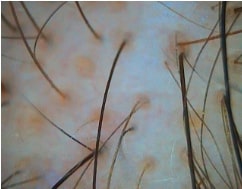
Fig. Androgenetic alopecia (yellow dots, anisotrichosis)

Fig. Alopecia areata (yellow dots, black dots)

Fig. Alopecia areata (exclamation mark hairs)
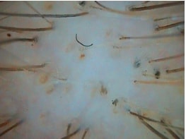
Fig. Trichotillomania