
What Skin Cancer Looks Like: Early Signs, Suspicious Spots and Changes to Watch For
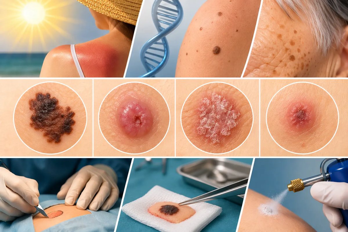
Changes in the skin are a natural part of life. With age, new moles, pigment spots, seborrhoeic keratoses and other benign growths appear. Many of them are harmless and do not need removing.
Sometimes, though, an ordinary-looking spot, a small bump or a sore that keeps crusting over turns out to be skin cancer. The difficulty is that a malignant lesion does not always hurt, itch or cause any other obvious symptoms.
That is why it helps to know what suspicious skin changes can look like, which signs to watch for and when it is time for dermoscopy.
What counts as a skin lesion?
A skin lesion is any growth that appears on the surface of the skin or within it. It can look like a mole, a spot, a bump, a growth or a small raised area. Lesions can be benign or malignant, and it is not always possible to tell which from appearance alone.
It is important to remember that not every new growth is a tumour, and not every tumour is malignant. Most skin lesions are benign.
A mole in a child, for example, may gradually get bigger as the body grows. That is not necessarily a sign of disease. A new spot, bump or sore in an adult that keeps changing, on the other hand, needs a careful look.
Do not try to work out what a lesion is just from its colour, size or shape. Benign and malignant lesions can look very similar.
Why does skin cancer so often go unnoticed?
One reason people see a doctor late is that there are no striking symptoms. Someone may ignore a small bump, a crust that keeps coming back or a slowly growing spot for years.
People often take such a change for an ordinary pimple, an age spot or the mark of a minor injury. If it does not hurt, it seems safe.
It also happens the other way round: a person comes to the doctor about a benign growth that gets in the way, gets injured or simply looks unattractive, and examining the rest of the skin reveals a suspicious lesion they never knew about.
That is why a skin check should not be limited to the one spot that worries you most, if the doctor thinks a wider examination is worthwhile.
Skin cancer does not always look obvious. At the same time, far from every change is malignant. The doctor's job is to separate what can be watched from what needs further tests or treatment.
Which skin changes should raise concern?
There are several signs worth paying attention to:
- a lesion is growing or changing shape;
- a mole changes colour or new shades appear;
- a lesion looks clearly different from the others;
- a crust keeps forming on the surface;
- a sore appears that does not heal;
- a lesion bleeds for no obvious reason or after a light touch;
- a persistent rough, scaly or thickened patch appears;
- new sensations appear: itching, pain or tenderness.
None of these signs on its own confirms skin cancer. But if a change persists, gets worse or keeps coming back, do not put off a check-up.
Pay particular attention to lesions that do not heal for a long time, crust over again after the crust falls off, or bleed from time to time.
The ugly duckling sign: a lesion that stands out
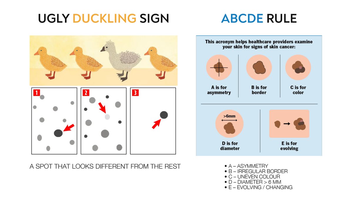
When you check your own skin, it helps to look not only at the features of each mole, but also at how it compares with the others on your body.
In dermatology this is called the ugly duckling sign. The idea is that a suspicious lesion often looks noticeably different from the rest of a person's moles.
Most of your moles may be small, evenly coloured and similar in shape. If one of them has an unusual colour, irregular edges or a different structure, it deserves special attention.
Being different does not necessarily mean being malignant. It is a signal that the lesion should be looked at more closely.
The ABCDE rule: how to assess suspicious moles
For a first impression of pigmented lesions, doctors use the ABCDE rule. It is an easy way to remember the main features that can point to melanoma.
- A for Asymmetry. One half of the mole differs from the other in shape or structure.
- B for Border. The edges are irregular, blurred or oddly shaped.
- C for Colour. The colour is uneven, with different shades of brown, black, red, pink, white or blue.
- D for Diameter. The lesion is larger than 6 mm across. Melanoma can be smaller, though, so a small size does not rule out danger.
- E for Evolving. The mole changes over time: it grows, changes shape, colour or structure, starts to bleed or causes new sensations.
Change over time matters most. If a lesion is changing, that is a reason to have it checked by a doctor, even if it is small or does not have every feature on the list.
The ABCDE rule is mainly a tool for spotting suspicious pigmented lesions, and it cannot rule out skin cancer if none of these features is present. Nor is it meant for diagnosing basal cell or squamous cell carcinoma yourself.
What does basal cell carcinoma look like?
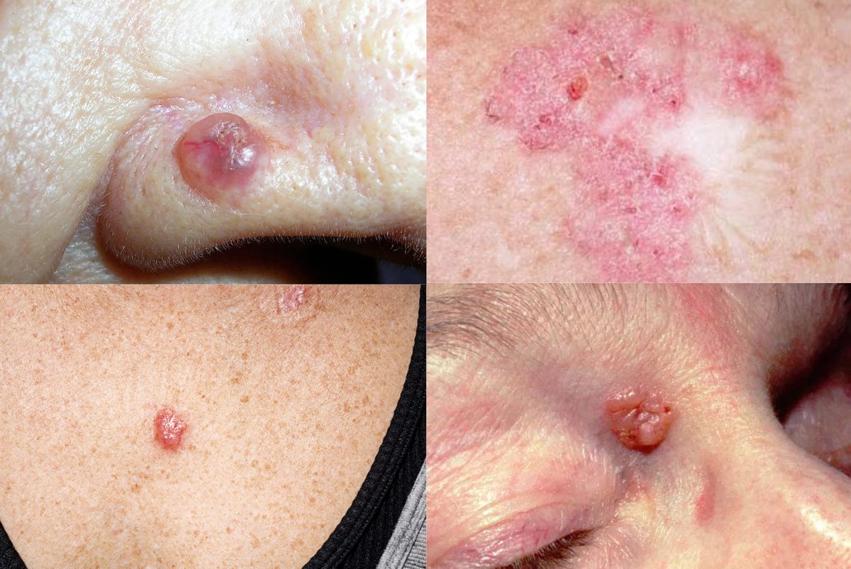
Basal cell carcinoma (BCC), also called basalioma, is one of the most common types of skin cancer. It usually grows slowly and rarely spreads, but without treatment it gradually destroys the surrounding tissue.
In the early stages, patients often describe it as a small pimple that will not go away.
BCC can look like:
- a shiny or pearly bump;
- a pink or reddish patch;
- a small growth with visible surface blood vessels;
- a sore that does not heal;
- an area that keeps forming a crust.
Over time an ulcer may form on the surface and bleed from time to time. The crust falls off, but the sore comes back in the same place.
These changes often appear on skin that gets a lot of sun: the face, nose, ears, neck and other exposed areas. BCC can, however, appear anywhere on the skin.
If a lesion is slowly growing, not healing or bleeding again and again, see a doctor. BCC cannot be confirmed by appearance alone: where indicated, histology is needed to make the diagnosis.
What does squamous cell carcinoma look like?
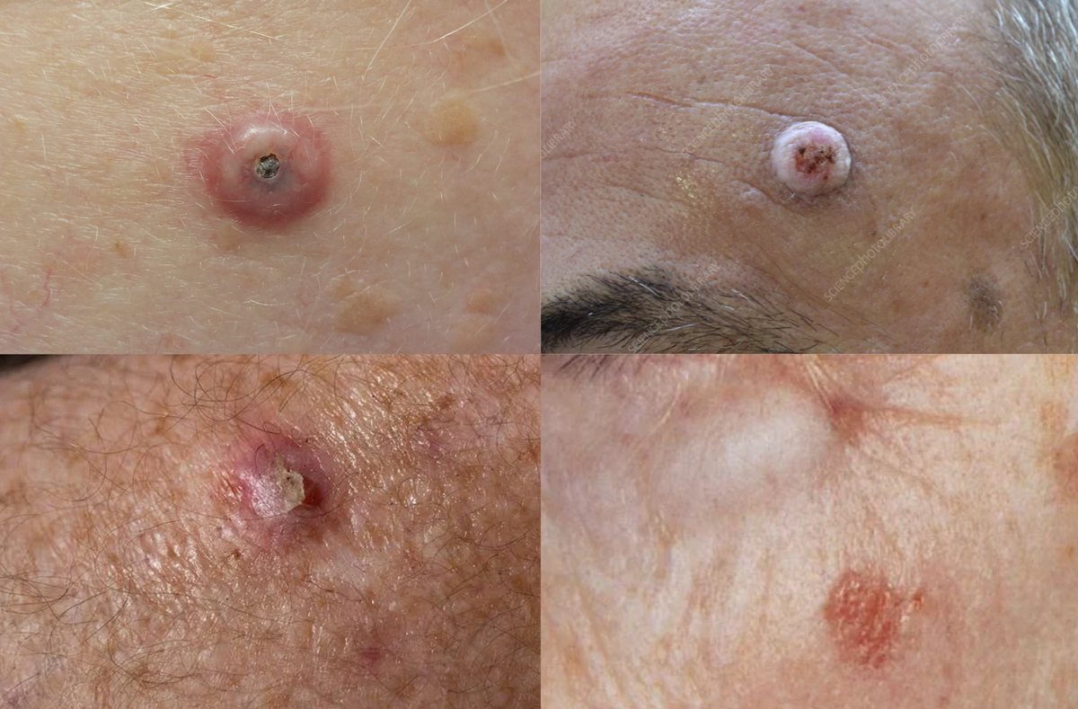
Squamous cell carcinoma (SCC) is another common type of non-melanoma skin cancer. Unlike many BCCs, in some cases it is more likely to spread to other tissues and metastasise. That is why suspicious lesions should be assessed without delay.
SCC can appear as:
- a rough, dry or scaly patch that does not go away;
- a firm red or skin-coloured bump;
- a crusted lesion or one with marked thickening of the top layer of skin;
- an ulcer that does not heal for a long time;
- an area that gradually grows, bleeds or crusts over again.
Sometimes the surface is heavily keratinised, with a crust or ulcer forming in the centre. Pain, tenderness or itching may appear in some cases, but their absence does not rule out the disease.
SCC is also more common on skin that has had long-term sun exposure, such as the face, ears, a scalp with thinning hair, the lips, the backs of the hands and other exposed areas.
Do not treat a persistent scaly patch, a firm lump or a sore that will not heal with ointments, and do not try to burn it off yourself. First find out what it is.
Can skin cancer look like an ordinary pimple or spot?
Yes. In the early stages some skin cancers can look like an ordinary pimple, irritation, a pigment spot or a small sore.
A pimple that will not go away does not by itself mean cancer, though. What matters is how long the lesion has been there, how it has changed, what it looks like and other clinical signs.
Lesions that need particular attention are those that:
- last longer than an ordinary inflamed spot would;
- are gradually getting bigger;
- regularly bleed or crust over;
- do not heal for no obvious reason;
- change colour, shape or structure;
- appear in someone who has had skin cancer before or has other significant risk factors.
Do not burn off such lesions, remove them with cosmetic products or use ointments on them without guidance. This can delay the correct diagnosis.
How to check your own skin
Self-examination helps you notice new lesions and changes in existing ones. Look not only at your face and hands, but also at the parts of your body that are harder to see.
During a skin check, look at:
- your face, ears and neck;
- your scalp;
- your back and the backs of your shoulders;
- your chest, stomach and sides;
- your arms, palms, fingers and nails;
- your legs, feet, soles and between your toes.
Some areas are hard to check on your own. If possible, ask someone close to you to look at your back and other spots you cannot see well.
It is useful to photograph lesions you are keeping an eye on, so you can notice changes. Photos do not replace dermoscopy or a professional assessment, however.
If you notice a suspicious change, do not wait for your next routine check-up.
Who should pay particular attention to skin cancer prevention?
Keeping a close eye on your skin is especially important if you have:
- a personal or family history of melanoma or other skin cancers;
- a large number of moles or atypical moles;
- fair skin that burns easily;
- a lot of accumulated sun exposure or a history of frequent sunburn;
- used tanning beds regularly in the past;
- a weakened immune system;
- had radiotherapy in the past or other established risk factors.
People in these groups may be advised to have individual monitoring, including dermoscopy and, where indicated, digital mole mapping.
There is no single check-up interval that suits everyone. How often you need one depends on your personal risk, history and earlier results.
Protecting your skin from too much UV matters too: use broad-spectrum sunscreen, wear covering clothes and a hat, seek shade and avoid tanning beds. Sun-safe habits are best formed in childhood.
Why dermoscopy is needed for diagnosis
An ordinary examination shows what a lesion looks like on the outside, but not always its inner structure. Dermoscopy magnifies the image and helps the doctor assess features that cannot be properly seen with the naked eye.
At the consultation the doctor asks when the lesion appeared and how it has changed, examines it and uses a dermatoscope to study its characteristic structures. Other areas of the skin are checked if needed.
The result helps decide what to do next: monitoring, further tests or removal with histology.
Dermoscopy is not a final diagnosis. If a malignant lesion is suspected, the doctor decides whether a tissue sample is needed for histology and which diagnostic method to use.
The main goal of a skin check is not to remove every lesion on the skin, but to find the ones that need special attention and not to miss dangerous changes.
When should you see a doctor?
Do not put off a consultation if a lesion is changing quickly, has an unusual colour or shape, bleeds regularly, crusts over or does not heal.
Do not wait for pain. Basal cell and squamous cell carcinomas can develop for a long time without noticeable symptoms.
At the same time, there is no need to keep looking for signs of disease or to treat every mole as dangerous. The sensible approach is to know your skin, notice changes and get a professional opinion in good time.
Skin lesion checks in Kharkiv
If you have noticed a new spot or bump, a mole that is changing or a sore that will not heal, have it examined by a dermato-oncologist.
At the consultation the doctor can perform dermoscopy, decide whether the lesion needs monitoring or further tests, and plan the next steps.
Not every lesion needs to be removed. But suspicious changes should not be ignored or dealt with on your own.
Book a consultation with dermoscopy in Kharkiv. A timely visit helps establish what a suspicious lesion is and choose a well-grounded plan for further tests or treatment.
Get a Consultation
Sources
- Centers for Disease Control and Prevention (CDC). Symptoms of Skin Cancer
- National Institute for Health and Care Excellence (NICE). Suspected cancer: recognition and referral
- European consensus-based interdisciplinary guideline for melanoma. Part 1: Diagnostics, Update 2024. PubMed
- American Academy of Dermatology (AAD). What to look for: ABCDEs of melanoma
- U.S. Preventive Services Task Force. Skin Cancer: Screening
About the author
Nazar V. Tsalin, MD, dermato-oncologist and surgical oncologist.
Focus: diagnosis and surgical treatment of skin lesions, dermoscopy, early detection of skin cancer.