
Mole Mapping in Kharkiv: Digital Total Body Mapping and Dermoscopy
What is mole mapping?
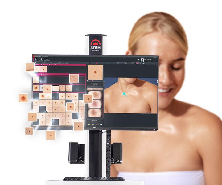
Mole mapping, or digital skin mapping, is an examination in which the whole surface of your skin is photographed, and then individual moles and other lesions that need watching are recorded close up with dermoscopy.
The point is not just to record what is on your skin today, but to be able to compare it over time. This matters most for people with many moles, who find it hard to keep track of every spot and to notice new or changing ones.
A new mole, or a change in an existing one, does not necessarily mean cancer. In some cases, though, such changes can be early signs of melanoma or another skin condition. That is why it is not enough to take photos: the results have to be read correctly.
Digital mapping lets the doctor see the overall picture of your skin, compare images between visits and decide which lesions need closer attention.
Who needs digital mole mapping?
Not everyone needs skin mapping. It is most useful for people with many melanocytic naevi (moles) and other factors that raise the risk of melanoma.
Digital mapping is worth considering if you have:
- a lot of moles that are hard to keep track of yourself;
- atypical moles with an unusual shape, uneven colour or irregular borders;
- had melanoma before;
- close relatives who have had melanoma, or known inherited risk factors;
- many moles combined with other risk factors;
- a dermatologist's advice to have long-term digital monitoring.
What counts is not only how many moles you have, but also their type, your medical history and other clinical factors.
According to the consensus recommendations of the International Dermoscopy Society, 60 or more melanocytic naevi are reason enough to consider digital monitoring. With more than 40, the decision depends on additional risk factors, such as previous melanoma, red hair and certain genetic traits.
These criteria help identify the people for whom mapping can be especially useful. They are not a universal rule, though, and they do not mean that mapping is wrong for everyone with fewer moles.
If you have only a few moles and no other risk factors, a standard dermatologist check with dermoscopy where needed is often enough.
How does skin mapping with FotoFinder work?
For digital mole checks our clinic uses the FotoFinder system, a combined hardware and software platform for photographic skin mapping and close-up dermoscopic records of individual lesions.
The examination has several steps.
1. Skin check and risk assessment.
Before the procedure the doctor goes over your medical history, looks at how many moles you have and what they are like, asks whether you or your family have had melanoma or other skin cancers, and decides whether digital mapping is indicated.
2. Photographing the body.
A series of standardised photos of different areas of the skin is taken. This records where the visible lesions are and creates your personal photo map.
3. Dermoscopy of individual moles.
Lesions that need a closer look are examined with a digital dermatoscope. The images are stored in the system for analysis and comparison later.
4. The doctor's assessment.
After the examination the doctor assesses how the lesions look, their dermoscopic features, and decides what to do next. Some moles may be marked for repeat monitoring, further tests or removal with histology.
Depending on the number of lesions and the scope of the check, the procedure usually takes about 30 to 45 minutes. It is non-invasive and painless.
How is digital skin mapping different from standard dermoscopy?
Standard dermoscopy lets the doctor examine individual moles and other skin lesions in detail. Digital mapping goes further: it photographs the whole surface of the skin and documents where the lesions that need watching are.
This gives the doctor a fuller picture of your skin and makes it possible to compare results over time. It matters most for people with many moles and other risk factors who need regular, systematic monitoring.
If you have only a few moles and other skin lesions, standard dermoscopy is often enough. If you have many, or there are reasons for long-term monitoring, digital mapping can tell the doctor more, because it records the state of all your skin and tracks changes over time.
So digital mapping does not replace standard dermoscopy. It extends what the examination can do, especially for people who need their skin lesions watched over time.
Why does it matter to watch moles over time?
At the first visit some lesions may show no clear signs of cancer and still need follow-up. In those cases the doctor may recommend repeat dermoscopy and comparison with the earlier images.
At the next mapping session the doctor checks whether the size, shape, colour or inner structure of the lesions has changed, and whether new spots have appeared that were not in the earlier photos.
The software helps store the images, compare them and flag areas that need a closer look. The final clinical assessment, and the decision on what to do next, is made by the doctor.
If a lesion has features suspicious for melanoma or another skin cancer, the necessary tests should not be put off just to keep watching it. In such cases the doctor decides whether removal or a biopsy with histology is needed.
Can artificial intelligence diagnose skin cancer on its own?
Modern digital dermoscopy systems may include software tools that analyse images, compare lesions and support the doctor's work. Some also use artificial intelligence algorithms.
Automated analysis, however, should not be taken as a final diagnosis in its own right. The images have to be read in a clinical context, taking into account your history, the features of the lesion and the findings of the examination.
The machine does not make the diagnosis. It helps the doctor gather and organise more information.
What really decides the quality of the examination is the experience of a specialist who is skilled in dermoscopy, can read what they see and can choose the right next step.
Can I get a copy of my mapping results?
Digital systems can store images and produce documentation of the examination. Where the feature is available, the images or a report can be exported as a PDF.
This can be useful if you plan to have future checks at another clinic. Earlier photos help the next doctor see the results of the previous examination and compare them with new findings.
How well the images can be compared depends on their quality, how complete the records are and whether the materials are compatible. So it is worth keeping your digital report and, if possible, bringing it to future appointments.
How often should mole mapping be done?
There is no single interval that suits everyone.
How often mapping is repeated depends on how many moles you have, your risk factors, the results of the previous examination and the features of the lesions being monitored.
For one person the doctor may recommend a planned follow-up after a set time, for another an earlier repeat visit. If a new suspicious change appears, there is no need to wait for the next scheduled mapping.
A repeat examination after one or two years may be right in some clinical situations, but it is not a universal recommendation for everyone.
Do I need skin mapping if I have fewer than 50 moles?
Not necessarily. The number of moles is only one of the criteria for choosing how to be examined.
If you have fewer than 50 moles, no significant risk factors and your moles look typically benign, the doctor may recommend a standard check with dermoscopy. In that situation mapping your whole body may not add enough to be necessary.
If you have atypical moles, have had melanoma, have a family history or other important risk factors, mapping can make sense even with fewer moles.
So there is no need to pick the most expensive or most complex option on your own. What matters is finding the method that best fits your own clinical situation.
Mole mapping and digital dermoscopy in Kharkiv
If you have many moles, atypical moles, or a doctor has advised watching your lesions over time, it is worth discussing digital skin mapping.
At the consultation the doctor will assess the number and features of your lesions, decide whether digital dermoscopy is indicated and help you choose the right kind of examination.
The aim is not to remove every mole, but to notice changes in time, find the lesions that need more attention and avoid unnecessary procedures.
Book a consultation for a mole check and digital skin mapping in Kharkiv. A professional assessment will show whether you need mapping, how often to come back and which lesions need further tests.
Get a Consultation
Scientific sources
- Russo T. et al. Indications for Digital Monitoring of Patients With Multiple Nevi: Recommendations from the International Dermoscopy Society. Dermatology Practical & Conceptual, 2022 (PubMed)
- NICE. Melanoma: assessment and management. Recommendations. Guidance on dermoscopy, photographic records and individual monitoring of suspicious pigmented lesions.
- Hobelsberger S. et al. Three-Dimensional Total Body Photography, Digital Dermoscopy, and in vivo Reflectance Confocal Microscopy for Follow-Up Assessments of High-Risk Patients for Melanoma: A Prospective, Controlled Study. Dermatology, 2024
- Ji-Xu A. et al. The Value of Total Body Photography for the Early Detection of Melanoma: A Systematic Review
About the author
Nazar V. Tsalin, MD, dermato-oncologist and surgical oncologist.
Focus: diagnosis and surgical treatment of skin lesions, dermoscopy, early detection of skin cancer.