Onychodystrophies: differential diagnostic markers
Ovcharenko Yu.S. Onychodystrophies: differential diagnostic markers // Дерматолог. – 2015. – Vol. 06, No. 01. – pp. 44–48 (original in Russian).
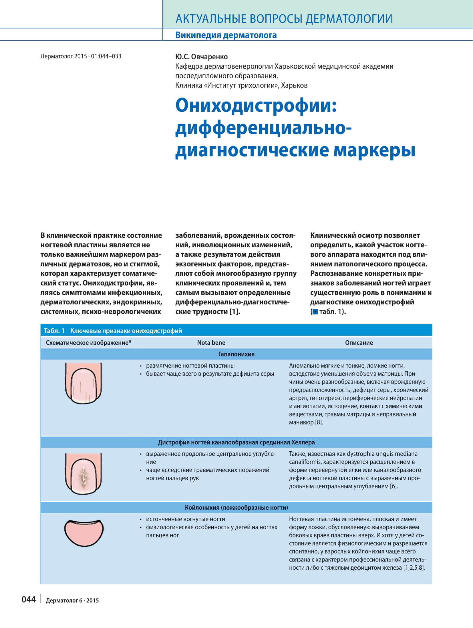
In clinical practice, the state of the nail plate is not only a key marker of various dermatoses but also a stigma that reflects a person’s somatic status. Onychodystrophies, as symptoms of infectious, dermatological, endocrine, systemic and psychoneurological diseases, congenital conditions and involutional changes, and as the result of exogenous factors, form a diverse group of clinical manifestations and therefore pose certain difficulties in differential diagnosis [1]. A clinical examination makes it possible to determine which part of the nail apparatus is affected by the pathological process. Recognising specific signs of nail disease plays an essential role in understanding and diagnosing onychodystrophies (Table 1).
Table 1. Key signs of onychodystrophies
Hapalonychia
- softening of the nail plate
- most often caused by sulphur deficiency
Abnormally soft, thin and brittle nails resulting from a reduced volume of the matrix. The causes are very diverse, including congenital predisposition, sulphur deficiency, chronic arthritis, hypothyroidism, peripheral neuropathies and angiopathies, emaciation, contact with chemicals, injuries to the matrix and improper manicure [8].
Heller’s median canaliform dystrophy
- a pronounced longitudinal central depression
- more often the result of traumatic damage to the fingernails
Also known as dystrophia unguis mediana canaliformis, it is characterised by splitting in the shape of an inverted fir tree or a canal-like defect of the nail plate with a pronounced longitudinal central depression [6].
Koilonychia (spoon nails)
- thinned, concave nails
- a physiological feature of children’s toenails
The nail plate is thinned and flat and has the shape of a spoon, because its lateral edges turn upwards. Although in children the condition is physiological and resolves spontaneously, in adults koilonychia is most often linked to the nature of their occupation or to severe iron deficiency [1,2,5,8].
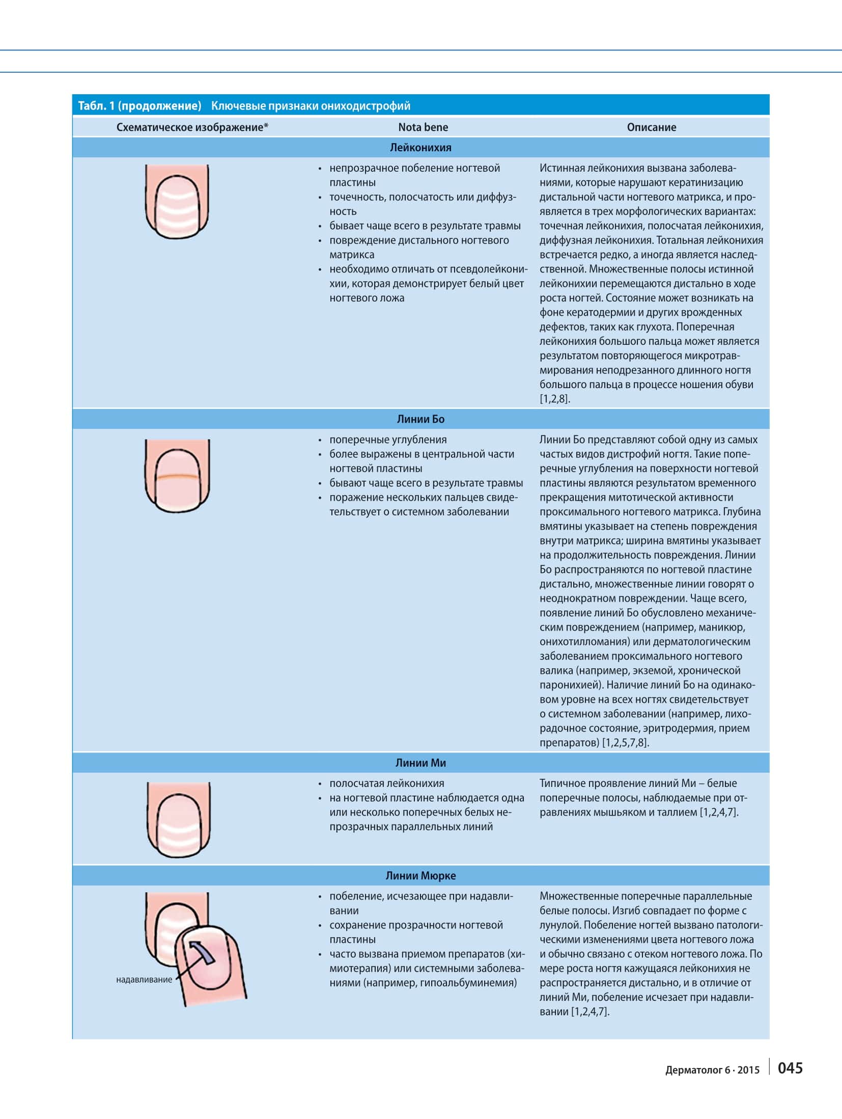
Leukonychia
- opaque whitening of the nail plate
- punctate, striate or diffuse
- most often caused by trauma
- damage to the distal nail matrix
- must be distinguished from pseudoleukonychia, in which the nail bed is white
True leukonychia is caused by diseases that disrupt keratinisation of the distal part of the nail matrix, and it occurs in three morphological variants: punctate leukonychia, striate leukonychia and diffuse leukonychia. Total leukonychia is rare and sometimes hereditary. The multiple bands of true leukonychia move distally as the nails grow. The condition may occur alongside keratoderma and other congenital defects such as deafness. Transverse leukonychia of the big toe may result from repeated microtrauma to an untrimmed long big toenail while wearing shoes [1,2,8].
Beau’s lines
- transverse depressions
- more pronounced in the central part of the nail plate
- most often caused by trauma
- involvement of several digits points to a systemic disease
Beau’s lines are one of the most common types of nail dystrophy. These transverse depressions on the surface of the nail plate result from a temporary arrest of mitotic activity in the proximal nail matrix. The depth of the dent indicates the degree of damage within the matrix; its width indicates how long the damage lasted. Beau’s lines move distally along the nail plate, and multiple lines indicate repeated damage. Most often, Beau’s lines are caused by mechanical damage (for example, manicure, onychotillomania) or by a dermatological disease of the proximal nail fold (for example, eczema, chronic paronychia). Beau’s lines at the same level on all nails point to a systemic disease (for example, a febrile state, erythroderma, medication) [1,2,5,7,8].
Mees’ lines
- striate leukonychia
- one or more transverse, white, opaque, parallel lines across the nail plate
The typical presentation of Mees’ lines is white transverse bands seen in arsenic and thallium poisoning [1,2,4,7].
Muehrcke’s lines
- whitening that disappears on pressure
- the nail plate stays transparent
- often caused by medication (chemotherapy) or systemic diseases (for example, hypoalbuminaemia)
Multiple transverse, parallel white bands. Their curve follows the shape of the lunula. The whitening is caused by pathological changes in the colour of the nail bed and is usually linked to oedema of the nail bed. As the nail grows, this apparent leukonychia does not move distally and, unlike Mees’ lines, the whitening disappears on pressure [1,2,4,7].
Micronychia
- a congenital condition
Small, short fingernails and toenails are often a congenital anomaly or are caused by diseases such as systemic scleroderma and trophoneuroses [8].
Onychauxis
- thickening of the nails
- subungual hyperkeratosis
- usually due to psoriasis or onychomycosis
Because of subungual shedding of the epidermis, the nail plate looks thickened. The causes of subungual hyperkeratosis include psoriasis, onychomycosis and eczema [1,2,8].
Onychogryphosis
- nails shaped like a ram’s horn
- a thick, hard, yellow-brown nail plate
- most often affects the big toenail
- elderly patients
Onychogryphosis is common in elderly people and almost exclusively affects the toenails, usually of the big toes. Because of asymmetric growth, the nail takes on the characteristic shape of a ram’s horn. The nail plate is thickened, hard and yellow-brown, often with multiple transverse grooves [1,2,8].
Onychocryptosis (ingrown nails)
- typically affects the proximal nail fold within the ventral nail plate and moves distally as the nails grow
- often bilateral
- associated with foot defects
- growth of granulation tissue
Traumatic damage to the toenails is most common in women who wear tight high-heeled shoes. Retronychia is caused by the nail growing into the proximal nail fold after trauma and may be accompanied by inflammation of the proximal nail fold. Chronic onychocryptosis is characterised by the growth of granulation tissue, which eventually becomes epithelialised [1,2,8].
Diagnosis and treatment at the Institute
Get a consultation
Onycholysis
- distal detachment of the nail plate
- the detached nail turns yellowish-white
- often results from external damage, psoriasis or onychomycosis
The nail plate separates from the nail bed; because air enters the subungual space, the plate looks white. Microbial colonisation or haemorrhage may cause pigmentation. Besides external damage, the diseases of the nail bed that most often lead to it are psoriasis and onychomycosis. UV exposure, alone or combined with drugs such as tetracycline, can trigger photo-onycholysis [1,2,6,7,8].
Idiopathic onycholysis
- fingernails only
- white or green nails
- no subungual hyperkeratosis
Idiopathic onycholysis is often the result of repeated exposure to water and irritants. The affected nail separates from the nail bed and often changes colour because of secondary microbial infection of the subungual space [1,2,7,8].
Onychomadesis
- proximal detachment of the nail
- most often caused by trauma
- involvement of several digits points to a systemic disease
The term onychomadesis describes the separation of the nail plate from the proximal nail fold associated with severe damage that leads to a complete arrest of nail matrix function. Onychomadesis appears as a groove that replaces the proximal nail plate. The causes of onychomadesis are the same as those of Beau’s lines [1,2,3,8].
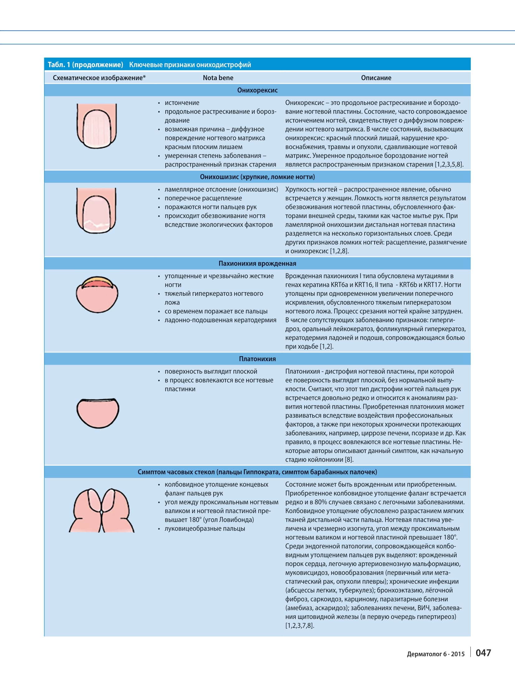
Onychorrhexis
- thinning
- longitudinal cracking and ridging
- a possible cause is diffuse damage to the nail matrix by lichen planus
- in a mild degree, a common sign of ageing
Onychorrhexis is longitudinal cracking and ridging of the nail plate. The condition, often accompanied by thinning of the nails, indicates diffuse damage to the nail matrix. Conditions that cause onychorrhexis include lichen planus, impaired blood supply, and injuries and tumours compressing the nail matrix. Mild longitudinal ridging of the nails is a common sign of ageing [1,2,3,5,8].
Onychoschizia (brittle, fragile nails)
- lamellar splitting (onychoschizia)
- transverse splitting
- affects the fingernails
- the nail dries out because of environmental factors
Brittle nails are common, usually in women. Fragility of the nail results from dehydration of the nail plate caused by environmental factors such as frequent hand washing. In lamellar onychoschizia the distal nail plate splits into several horizontal layers. Other signs of brittle nails include splitting, softening and onychorrhexis [1,2,8].
Pachyonychia congenita
- thickened and extremely hard nails
- severe hyperkeratosis of the nail bed
- over time affects all digits
- palmoplantar keratoderma
Pachyonychia congenita type I is caused by mutations in the keratin genes KRT6a and KRT16, type II by mutations in KRT6b and KRT17. The nails are thickened, with increased transverse curvature caused by severe hyperkeratosis of the nail bed. Cutting the nails is extremely difficult. Associated signs include hyperhidrosis, oral leukokeratosis, follicular hyperkeratosis, and keratoderma of the palms and soles with pain on walking [1,2].
Platonychia
- the surface looks flat
- all nail plates are involved
Platonychia is a dystrophy of the nail plate in which its surface looks flat, without the normal convexity. This type of fingernail dystrophy is thought to be fairly rare and is classed among developmental anomalies of the nail plate. Acquired platonychia may develop under occupational factors and in some chronic diseases, for example cirrhosis of the liver, psoriasis and others. As a rule, all nail plates are involved. Some authors describe this sign as the initial stage of koilonychia [8].
Watch-glass nails (Hippocratic fingers, drumstick fingers)
- club-shaped thickening of the terminal phalanges of the fingers
- the angle between the proximal nail fold and the nail plate exceeds 180° (Lovibond’s angle)
- bulbous fingers
The condition may be congenital or acquired. Acquired clubbing of the phalanges is rare and in 80% of cases is linked to lung disease. Clubbing results from overgrowth of the soft tissues of the distal part of the finger. The nail plate is enlarged and excessively curved, and the angle between the proximal nail fold and the nail plate exceeds 180°. Endogenous conditions accompanied by clubbing of the fingers include congenital heart disease, pulmonary arteriovenous malformation, cystic fibrosis, neoplasms (primary or metastatic cancer, pleural tumours); chronic infections (lung abscesses, tuberculosis); bronchiectasis, pulmonary fibrosis, sarcoidosis, carcinoma, parasitic diseases (amoebiasis, ascariasis); liver disease, HIV, and thyroid disease (above all hyperthyroidism) [1,2,3,7,8].
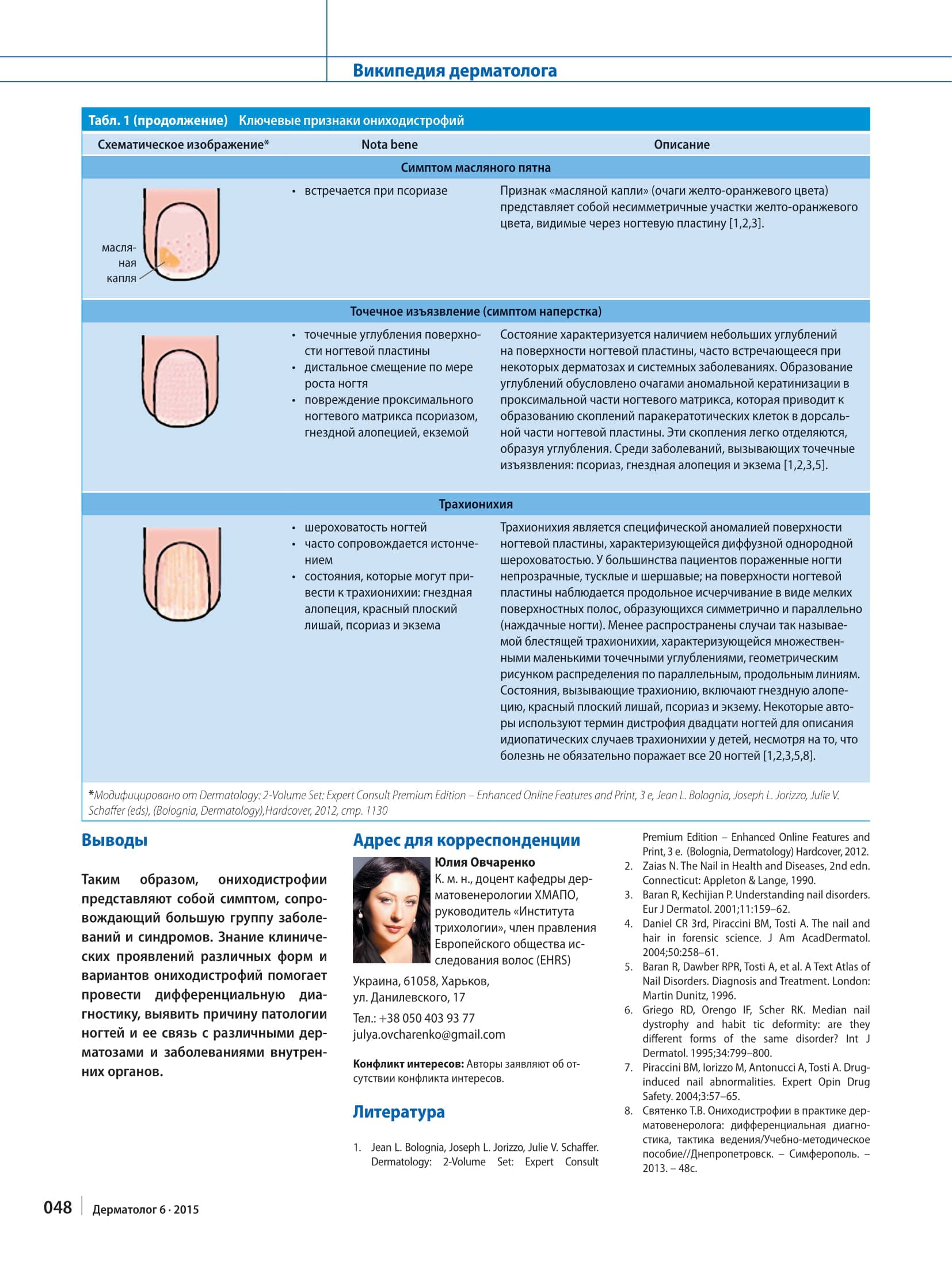
Oil spot sign
- seen in psoriasis
The “oil drop” sign (yellow-orange patches) consists of asymmetric yellow-orange areas visible through the nail plate [1,2,3].
Pitting (thimble sign)
- pinpoint depressions in the surface of the nail plate
- moves distally as the nail grows
- damage to the proximal nail matrix by psoriasis, alopecia areata or eczema
The condition is characterised by small depressions in the surface of the nail plate and is common in some dermatoses and systemic diseases. The pits are caused by foci of abnormal keratinisation in the proximal part of the nail matrix, which lead to clusters of parakeratotic cells in the dorsal part of the nail plate. These clusters come away easily and leave pits. Diseases that cause pitting include psoriasis, alopecia areata and eczema [1,2,3,5].
Trachyonychia
- rough nails
- often accompanied by thinning
- conditions that can lead to trachyonychia: alopecia areata, lichen planus, psoriasis and eczema
Trachyonychia is a specific abnormality of the nail plate surface characterised by diffuse, uniform roughness. In most patients the affected nails are opaque, dull and rough; the nail plate surface shows longitudinal striation in the form of fine superficial ridges arranged symmetrically and in parallel (“sandpaper nails”). Less common is so-called shiny trachyonychia, characterised by multiple small pits arranged in a geometric pattern along parallel longitudinal lines. Conditions that cause trachyonychia include alopecia areata, lichen planus, psoriasis and eczema. Some authors use the term twenty-nail dystrophy for idiopathic cases of trachyonychia in children, even though the disease does not necessarily affect all 20 nails [1,2,3,5,8].
*The schematic drawings of each sign (on the original publication pages below) are modified from: Dermatology: 2-Volume Set: Expert Consult Premium Edition – Enhanced Online Features and Print, 3 e, Jean L. Bolognia, Joseph L. Jorizzo, Julie V. Schaffer (eds), (Bolognia, Dermatology), Hardcover, 2012, p. 1130.
Conclusions
Onychodystrophies are thus a symptom that accompanies a large group of diseases and syndromes. Knowing the clinical presentation of the different forms and variants of onychodystrophy helps to make a differential diagnosis and to identify the cause of nail pathology and its connection with various dermatoses and diseases of the internal organs.
Conflict of interest: the authors declare no conflict of interest.
References
- Jean L. Bolognia, Joseph L. Jorizzo, Julie V. Schaffer. Dermatology: 2-Volume Set: Expert Consult Premium Edition – Enhanced Online Features and Print, 3 e. (Bolognia, Dermatology) Hardcover, 2012.
- Zaias N. The Nail in Health and Diseases, 2nd edn. Connecticut: Appleton & Lange, 1990.
- Baran R, Kechijian P. Understanding nail disorders. Eur J Dermatol. 2001;11:159–62.
- Daniel CR 3rd, Piraccini BM, Tosti A. The nail and hair in forensic science. J Am AcadDermatol. 2004;50:258–61.
- Baran R, Dawber RPR, Tosti A, et al. A Text Atlas of Nail Disorders. Diagnosis and Treatment. London: Martin Dunitz, 1996.
- Griego RD, Orengo IF, Scher RK. Median nail dystrophy and habit tic deformity: are they different forms of the same disorder? Int J Dermatol. 1995;34:799–800.
- Piraccini BM, Iorizzo M, Antonucci A, Tosti A. Drug-induced nail abnormalities. Expert Opin Drug Safety. 2004;3:57–65.
- Святенко Т.В. Ониходистрофии в практике дерматовенеролога: дифференциальная диагностика, тактика ведения/Учебно-методическое пособие//Днепропетровск. – Симферополь. – 2013. – 48с.
Original publication pages