
Basal Cell Carcinoma Removal in Kharkiv: Surgical Treatment Options
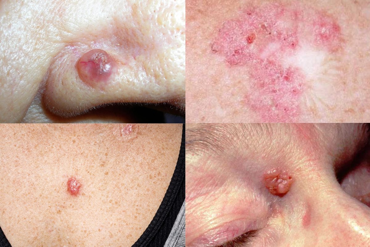
Basal cell carcinoma (BCC), also called basalioma, is a malignant skin tumour. It usually appears on skin that gets the most sun: the face, nose, ears, neck and scalp. It can, however, develop anywhere on the body.
BCC tends to grow slowly and very rarely spreads to distant organs. That does not make it harmless. Left untreated, the tumour gradually destroys the surrounding tissue, grows larger and makes later surgery more difficult.
Basal cell carcinoma removal in Kharkiv starts with a careful assessment of the lesion using modern methods, above all dermoscopy. The diagnosis is confirmed with a biopsy and histology, and the treatment is chosen according to the tumour's location, size and histological type.
What is basal cell carcinoma and what causes it?
BCC is one of the most common types of skin cancer. It arises from cells of the epidermis or related structures and grows mainly locally, invading the tissue around it.

The main risk factor is ultraviolet (UV) radiation. It affects skin cells throughout life, and the DNA damage builds up over time. Other factors also matter:
- frequent sunburns in the past;
- long hours in the sun without proper protection;
- tanning beds;
- fair skin;
- older age;
- a weakened immune system (immunosuppression);
- certain inherited syndromes and a family history of skin cancer.
Sun protection matters most for people who spend a lot of time outdoors for work, including military personnel, construction workers and anyone else regularly exposed to direct sunlight.
What does basal cell carcinoma look like?
BCC can look very different from one person to another. Sometimes it resembles an ordinary pimple that will not go away, a small sore that keeps crusting over, or a pink scaly patch.
The most common clinical form is nodular BCC, and it is the clearest example of how the tumour develops.
Nodular BCC
At first, a small skin-coloured, pink or pearly bump usually appears. Tiny blood vessels may be visible on its surface.
Over time the bump slowly grows. An erosion or ulcer may form in the centre, bleeding from time to time and crusting over. Sometimes the lesion stays barely noticeable and painless for a long time, which is why people often put off seeing a doctor.
Superficial BCC
It can look like a flat pink or reddish scaly patch and is sometimes mistaken for dermatitis or another inflammatory skin condition. It is more common on the trunk.
Sclerosing (morpheaform) BCC
This form can look like a pale, scar-like area with blurred edges. It can spread deeper and further than the visible changes suggest, so it needs especially careful diagnosis and treatment planning.
There are other histological subtypes of BCC as well, which differ in how they grow and how likely they are to recur. The final treatment plan depends not only on how the lesion looks, but also on other clinical factors and, where indicated, on the histology results.
Why is basal cell carcinoma dangerous?
The main danger of BCC is that it gradually destroys tissue where it grows.
Unlike many other cancers, basal cell carcinoma very rarely metastasises. But if it is left untreated for a long time, it can invade the deeper layers of the skin, the fat beneath it, cartilage, muscle and even bone.
Cases are especially challenging when the tumour sits on the nose, eyelids, ears or other areas where removing even a small amount of tissue can affect how the area works or looks.
The longer the tumour goes untreated, the more tissue may need to be removed. This can make reconstruction harder and leave a larger scar.
Metastatic BCC is exceptionally rare. Its rarity, however, is no reason to delay treatment.
How basal cell carcinoma is diagnosed
The examination starts with a consultation with a doctor and an assessment of the skin lesion. The goal is not only to see whether it shows signs of malignancy, but also to weigh the factors that will shape the choice of treatment.
1. Medical history
The doctor asks when the lesion appeared, how it has changed, and whether it has bled, ulcerated or crusted over repeatedly. Previous skin cancers, sun exposure and family history are also taken into account.
2. Skin examination
The doctor assesses the size, location, borders and appearance of the lesion. If indicated, they may recommend a full-body skin check, since one patient can have several BCCs or other lesions that need attention at the same time.
3. Dermoscopy
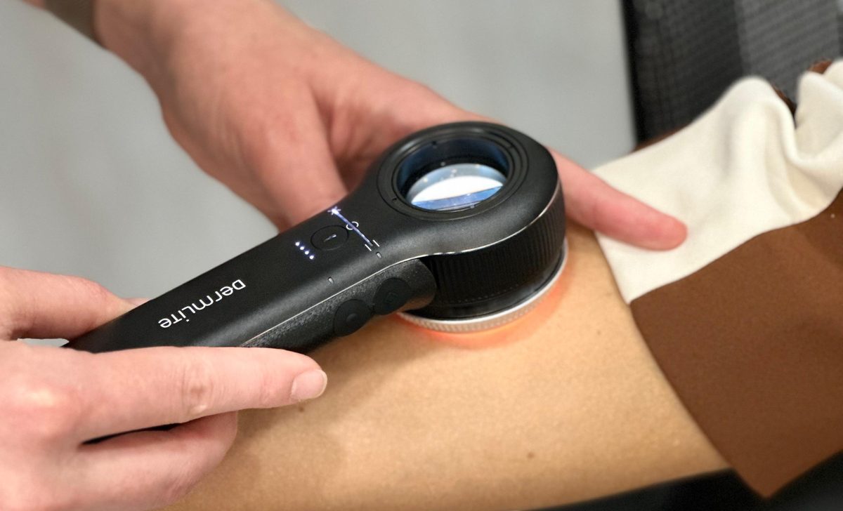
Dermoscopy is a non-invasive way of examining the skin with a special optical device, the dermatoscope. It reveals structures that cannot be properly assessed with the naked eye.
During dermoscopy the doctor looks for structures that help tell BCC apart from benign lesions and from some other skin cancers.
Dermoscopy cannot always confirm the diagnosis on its own, though. If the clinical picture is unclear or the lesion shows signs of a tumour, histological confirmation is needed.
4. Biopsy and histology
A biopsy means taking a tissue sample to examine under a microscope. It is usually done under local anaesthetic. One option is a punch biopsy, where a special instrument removes a small cylinder of tissue.
In some cases other types of biopsy are used, or the lesion is removed surgically straight away and the whole specimen is sent for histology.
Histology confirms the diagnosis and identifies the tumour type. After a standard surgical excision it also shows whether the margins of the removed tissue are clear. Standard histology, however, does not map the margins as completely as Mohs micrographic surgery does.
5. Is a CT or MRI scan needed?
Computed tomography and magnetic resonance imaging are not required for every patient with BCC.
How is basal cell carcinoma treated?
Treatment depends on the size and location of the tumour, its histological type, how clearly its borders are defined, any previous treatment, the state of the patient's immune system and other risk factors.
For most BCCs the main treatment is complete surgical removal. Other methods can be used in suitable clinical situations, but they are not right for every tumour.
Surgical removal of BCC
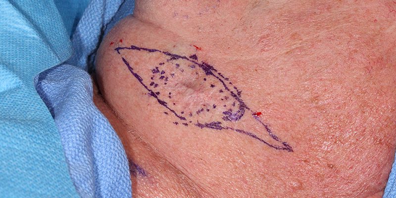
In a standard surgical excision the doctor removes the tumour with a set margin of healthy skin around its visible edges. The width of the margin depends on the features of the lesion and the risk of recurrence.
The wound is then closed with stitches, using a local skin flap if needed, and the removed tissue is sent for histology.
This method is often used for BCCs with well-defined borders and a low risk of recurrence, when an adequate margin can be taken without noticeably affecting function or appearance. Most often this is on the trunk, where there is enough spare tissue.
If histology finds tumour cells at the edge of the removed tissue, a second procedure may be needed to remove the tumour completely.
Mohs micrographic surgery
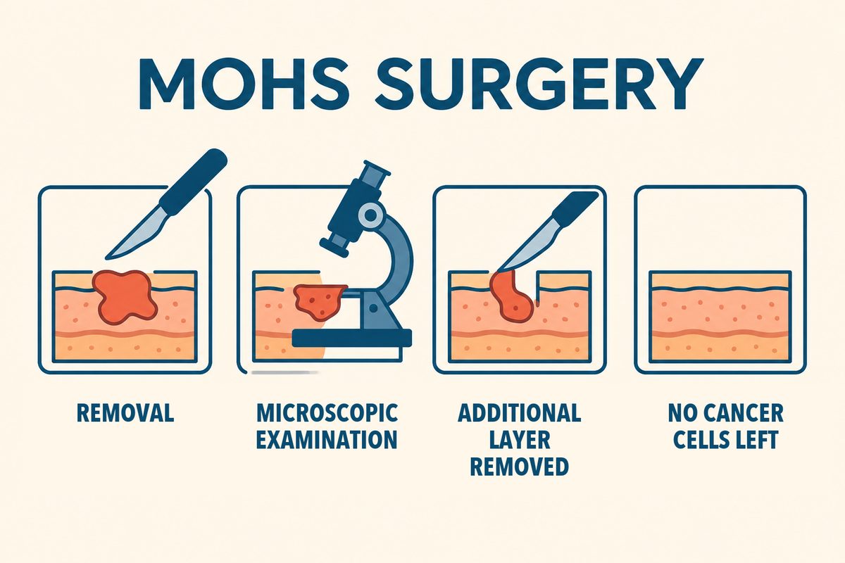
Mohs surgery removes the tumour step by step while checking its margins under a microscope. It is especially valuable when as much healthy tissue as possible must be preserved, for example on the nose, eyelids, lips and other anatomically delicate areas.
The tumour is removed in successive stages. Each layer is mapped and examined under the microscope using a special technique. If tumour cells remain at any point along the margin, an additional layer is removed from that exact spot.
The process is repeated until the margins are clear. This makes it possible to control both the side and deep margins of the excision while sparing as much healthy tissue as possible.
In many cases the operation is done under local anaesthetic. Once the tumour is removed and the wound is reconstructed, the patient can usually go home the same day if their condition allows.
How does Mohs surgery differ from standard excision?
In a standard excision the tumour is removed in one stage, and the final histology result comes later. If tumour remnants are found, a repeat excision may be needed.
In Mohs surgery the tissue is examined stage by stage, and the wound is closed only once the margins are clear. This gives better control over complete removal and spares more healthy tissue.
Not every patient needs Mohs surgery. Whether it makes sense depends on the risk of recurrence, the location and the features of the tumour. In suitable patients it achieves very high tumour control rates (99%), but no method can guarantee that a tumour will never come back.
Topical treatment with imiquimod
Imiquimod 5% cream works by stimulating the local immune response. It may be prescribed for some low-risk superficial BCCs when this approach suits the clinical situation.
The treatment takes a long time and can cause redness, irritation, crusting and other local reactions. Its success depends on following the prescribed regimen closely and on whether this treatment is right for the particular patient.
Imiquimod is not a universal substitute for surgery. It does not work for every type of BCC, and when a lesion disappears from view, that does not always mean all the tumour cells are gone.
Cryosurgery
Cryosurgery destroys tumour tissue with extreme cold, usually liquid nitrogen.
It may be considered for some small, low-risk BCCs if the tumour's characteristics allow it. Afterwards a blister, crusting, changes in pigmentation and a scar are possible.
An important limitation of cryosurgery is that it does not allow full histological control of the margins. As a result, the risk of incomplete treatment and recurrence may be higher than after appropriate surgery.
Electrosurgery and laser treatment
Some destructive methods can be used in selected cases of low-risk BCC. They should not, however, be regarded as an equal alternative to standard excision or Mohs surgery for every skin cancer.
When tissue is destroyed, it is impossible to map all the margins histologically the way it is done in micrographic surgery. That is why these methods are chosen only after careful assessment of their indications and limitations.
Why you should not put off treatment
BCC can go a long time without pain or other obvious symptoms. But slow growth does not mean the tumour will go away on its own.
Trying to burn off, cut off or treat the lesion yourself with destructive products can damage the tissue and delay the correct diagnosis. If a lesion looks smaller or its surface heals for a while, that does not prove the tumour is gone.
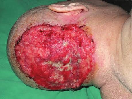
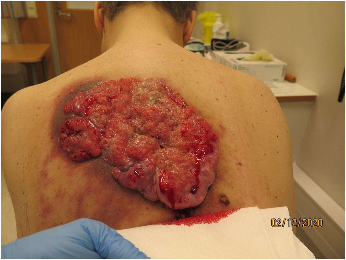
Seeing a doctor early makes it possible to assess the lesion, order the right tests and choose a treatment that takes the risks into account. Follow-up checks after treatment matter too, because people who have had BCC can develop a new skin cancer or a recurrence of the old one.
Basal cell carcinoma removal in Kharkiv
If you have a spot that has not healed for a long time, bleeds now and then, is growing or changing, have it checked by a doctor.
At the consultation the doctor can assess the lesion, perform dermoscopy and decide whether a biopsy or other tests are needed. If BCC is confirmed, the doctor will help you choose the best treatment.
The removal method is chosen individually, taking into account the size and location of the tumour, its histological features and the need to preserve as much healthy tissue as possible.
Do not delay checking a suspicious lesion. Timely diagnosis and the right treatment help bring the tumour fully under control and reduce the risk of serious damage to the surrounding tissue.
Get a Consultation
Medical sources
- European consensus-based interdisciplinary guideline for diagnosis and treatment of basal cell carcinoma, update 2023. PubMed
- NCCN Guidelines for Patients: Basal Cell Skin Cancer
- American Academy of Dermatology: Basal cell carcinoma, diagnosis and treatment
About the author
Nazar V. Tsalin, MD, dermato-oncologist and surgical oncologist.
Focus: diagnosis and surgical treatment of skin lesions, dermoscopy, early detection of skin cancer.