
Myths About Moles and Skin Cancer: What Patients Fear and What You Must Never Do
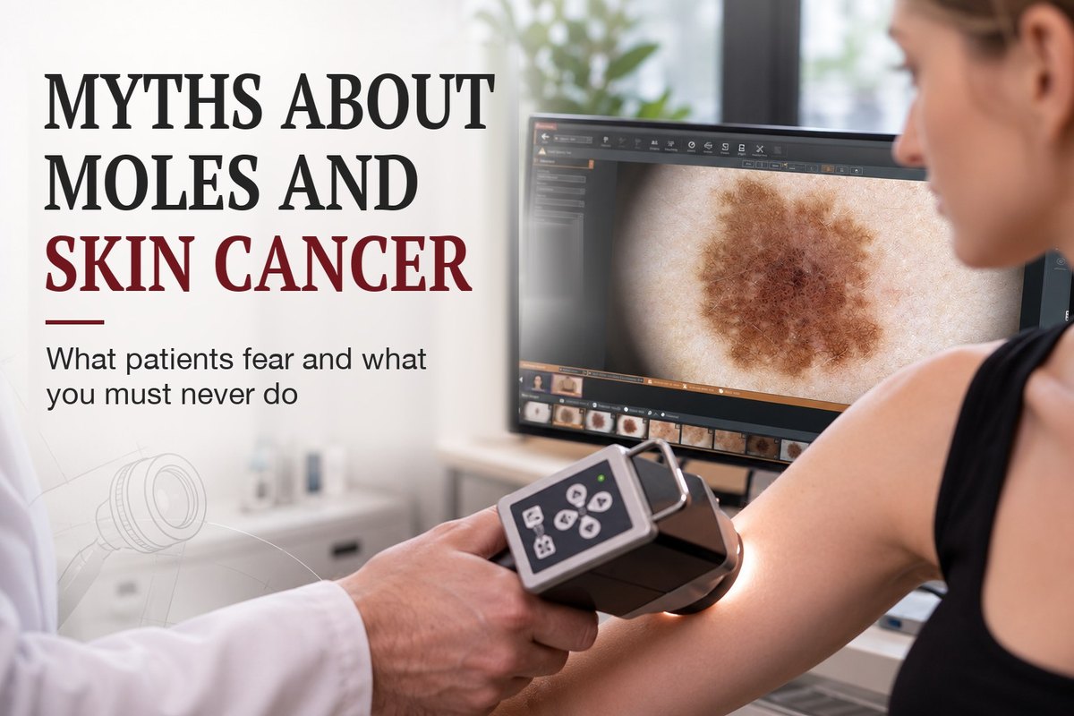
Introduction: when fear is more dangerous than the check-up
Most people have moles. Some stay the same for years, others change or appear with age. But when you notice an unusual spot, fear and worry often follow: "What if it's cancer?", "If I have it removed, will it spread?", "Maybe it's better to leave it alone?".
Because of these fears, people sometimes put off seeing a doctor, try to judge the spot from photos online or use home removal methods.
In dermato-oncology one thing matters: not every mole is dangerous, but every suspicious lesion needs a proper assessment. Fear does not make a diagnosis, and self-treatment can cause complications and delay the tests you need.
This article looks at common myths about moles and skin cancer, what is really worth worrying about and what you should never do.
Myth 1. If a mole doesn't hurt, it's safe
This is one of the most common beliefs, and one of the most dangerous.
In the early stages, melanoma and other skin cancers may cause no pain, itching or any other noticeable symptoms. A person can ignore a slowly changing spot for years.
A mole should be judged by more than how it feels. Important signs are:
- a change in size or shape;
- new colours or uneven colouring;
- asymmetry or irregular borders;
- a spot that looks clearly different from the others;
- bleeding, crusting or a surface that won't heal;
- new or growing changes that persist over time.
For a first check, even at home, I recommend the "ugly duckling" rule: look for the spot, or several spots, that stand out and don't match the rest.
Or the ABCDE rule: asymmetry, border irregularity, colour variation, diameter and, as one of the key criteria, evolution, meaning changes in the spot over time.
Still, this rule cannot rule out melanoma on its own. Small spots can be suspicious too, and some types of melanoma lack the typical dark colour.
No pain is no proof that a spot is benign. If a mole is changing or you are unsure about it, see a doctor.
Myth 2. It's better not to remove a mole, or the cancer will spread or it will turn into melanoma
Fear of mole removal still leads many people to turn down the examination and treatment they need. Some are convinced that any procedure on a skin lesion can trigger cancer or make a tumour spread.
Modern medicine does not support the idea that a properly performed biopsy or surgical removal of a suspicious lesion causes cancer to spread. On the contrary, the right diagnostic procedure is a key step in making the diagnosis and planning treatment.
The real problem is usually different: the patient already has a skin cancer but doesn't know it, because it has not been properly diagnosed. Sometimes such lesions are removed in beauty salons with lasers or other destructive methods that leave no proper tissue for histology. As a result, the diagnosis comes late.
That is why, before any removal, the lesion has to be assessed properly, the reasons for the procedure confirmed and a method chosen that will show what the lesion actually is.
When melanoma is suspected, doctors usually prefer a complete diagnostic excision with a small clinical margin, typically about 1 to 3 mm, where this is technically possible. In some cases, for example with large lesions or in difficult anatomical areas, other biopsy methods may be used.
It is important to understand that a diagnostic excision and the definitive removal of a confirmed melanoma are different stages of treatment. Once the diagnosis is made, the extent of the final surgery is decided according to the tumour's histological features and current clinical guidelines. An unnecessarily wide first excision of a suspicious mole does not improve the diagnosis, but it can enlarge the tissue defect and the scar and seriously reduce the diagnostic value of the procedure for the next stages of treatment.
So I advise patients to see a doctor who specialises in diagnosing and treating skin lesions. Don't hesitate to ask about the medical training and qualifications of whoever performs the procedure. It is your right and an important part of safe treatment.
A suspicious mole must never be cut off at home, burned with celandine or other harsh products, or removed in a beauty salon without a proper diagnostic assessment. The method should be chosen by a doctor, based on the clinical situation and the need for histopathology.
Don't put off an examination because you are afraid of removal. It is far more important to find out in time what has actually appeared on your skin. Uncertainty and delayed diagnosis are what can cost the time needed for timely treatment.
Myth 3. Once a lesion is removed, histology isn't needed
How a mole looks, even under dermoscopy, does not always allow a final diagnosis.
Histology means examining the tissue under a microscope, and in many cases it is the key step in confirming or ruling out melanoma. It also shows tumour features that matter for further treatment.
If a lesion was removed for diagnosis or cancer is suspected, the tissue must be sent for proper histopathology in line with the clinical situation.
Don't consider the problem solved just because the spot has physically gone from your skin. What matters is knowing exactly what was removed.
Myth 4. If a mole looks like a picture online, the diagnosis is obvious
Photos online can help you understand which skin changes should worry you. But they cannot replace a clinical examination and professional diagnosis.
The same goes for apps and artificial intelligence systems. They can mistake a skin cancer for a benign lesion or, the other way round, flag a benign lesion as suspicious. So the result of an automated analysis is never a final diagnosis.
Photo quality depends on lighting, focus, angle, camera and scale. Besides, patients usually can't take a dermoscopic image themselves, with the magnification and detail a professional assessment needs.
Another problem is that different benign and malignant lesions can look alike. Patients often call every spot on their skin a mole, while in fact it may be a seborrhoeic keratosis, an angioma, a naevus, basal cell carcinoma, melanoma or another condition.
For example, an angioma is a vascular growth of the skin, not in itself a sign of liver disease. And a spot that looks like an ordinary mole is not always a melanocytic naevus. Its nature often cannot be judged from an everyday description or a photo.
At a consultation the doctor looks not only at the colour and shape of the lesion, but also at your history, how it has changed, where it is, its dermoscopic features and other clinical data. Further tests or histology are ordered if needed.
A photo can be a reason to book a consultation, but it is no proof that a lesion is benign. If a spot is changing, bleeding, looks unusual or worries you, the right decision is to see a dermato-oncologist, not to diagnose yourself from pictures online.
Myth 5. Only older people get skin cancer
The risk of many skin cancers rises with age, but melanoma can also develop in young people.
Many factors affect the risk: UV exposure, past sunburns, the number and type of moles, a personal or family history of melanoma, inherited predisposition and certain immune system conditions.
Being young does not rule out cancer. Likewise, having many moles does not mean each of them is dangerous.
What matters is assessing your own risk factors and watching for new or changing spots at any age.
Myth 6. If a spot is small, it can't be melanoma
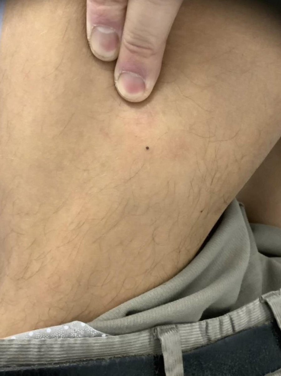
The photo shows a patient with melanoma in situ on the lateral surface of the left thigh.
Size is only one of the criteria. Melanoma can be small, especially at an early stage.
Just as important are changes over time, asymmetry, colour, borders and dermoscopic structure. Some lesions need assessing even when they are smaller than 6 mm across.
Don't wait until a mole gets bigger, starts to hurt or bleeds. If it is changing, it is better to have it checked sooner.
Myth 7. If my doctor recommends a biopsy, it means I have cancer
A recommendation for a biopsy is not a cancer diagnosis.
A biopsy is done precisely to obtain tissue and make an accurate diagnosis when a clinical examination is not enough or the lesion has suspicious features.
The result may confirm a benign lesion, show precancerous changes or establish a cancer diagnosis. What happens next depends on the result.
A referral for a biopsy is not a verdict. It is a diagnostic step that helps choose the right course of action.
What you must never do with a suspicious skin lesion
Some actions can injure a lesion, make it harder to assess or delay the tests you need.
1. Don't remove moles yourself.
Never cut them off with a blade or scissors, tie them off with thread, prick them or try to pull them out. This can cause bleeding, infection, incomplete removal and scarring. It also makes a proper diagnosis impossible.
2. Don't burn lesions with acids, alkalis or home remedies.
Wart removers, concentrated acids and other harsh substances can cause chemical burns and damage the tissue. If the lesion turns out to be cancer, self-treatment can delay the right diagnosis.
3. Don't treat a suspicious mole with creams instead of getting it checked.
No cream can reliably rule out melanoma based on how a lesion looks. Less inflammation for a while, or a change in the surface, does not prove the problem has gone.
4. Don't put off a consultation for fear of hearing a diagnosis.
If a lesion is changing, bleeding, ulcerating or not healing, don't wait for months hoping it will go away on its own. A timely examination shows whether further tests are needed.
5. Don't remove a suspicious lesion for cosmetic reasons alone without a proper assessment.
Before any procedure it is important to check for signs of cancer and decide which removal method is appropriate. If melanoma is suspected, the plan must allow for full histopathology.
6. Don't refuse histology when it is indicated.
Only examining the tissue can confirm the diagnosis and show the tumour features needed for further treatment.
7. Don't treat a normal photo or a past examination as a guarantee for the future.
If a lesion keeps changing after the consultation or new symptoms appear, see a doctor again. An earlier benign finding does not rule out a new lesion or new changes.
When should you see a dermato-oncologist?
Book a check if you notice:
- a new mole or other unusual spot that looks different from the rest;
- a change in the size, shape or colour of an existing mole;
- irregular borders or uneven colouring;
- bleeding for no obvious reason;
- a crust that keeps coming back, an ulcer or a sore that won't heal;
- a new dark streak under a nail with no clear cause;
- a lesion that changes quickly or keeps worrying you.
Not all of these signs mean cancer. But they are a reason for a professional assessment. If melanoma or another cancer is suspected, the doctor will decide whether a biopsy or another diagnostic step is needed.
How to look after your skin sensibly
Preventing skin cancer does not mean constant fear of every mole or removing every spot. It means paying attention to changes, protecting yourself from too much UV and seeing a doctor when there is a reason to.
There is no need to check every patch of skin anxiously every day or to diagnose yourself from photos. It is more useful to examine your skin from time to time, know your moles and get help if something changes.
For people at higher risk of melanoma, the doctor may recommend regular check-ups, dermoscopy or digital skin mapping. How often depends on the individual.
The main thing is not to ignore suspicious changes, but also not to conclude it is cancer without a proper diagnosis.
Conclusion
Fear of skin cancer is understandable, but it should never be a reason to skip an examination. Not every mole is dangerous, an accidental injury does not mean melanoma will develop, and a recommended biopsy is not a cancer diagnosis.
The real dangers are removing suspicious lesions yourself, burning them with unknown products, ignoring changes and putting off the diagnosis you need.
If a mole is changing, bleeding or looks unusual, don't try to treat it yourself. See a dermato-oncologist, who will decide what to do next based on an examination and the tests you need.
Get a Consultation
Scientific sources
- Amaral T. et al. Cutaneous melanoma: ESMO Clinical Practice Guideline for diagnosis, treatment and follow-up. Annals of Oncology. Guideline update, November 2024 (full text)
- Garbe C. et al. European consensus-based interdisciplinary guideline for melanoma. Part 1: Diagnostics, Update 2024. European Journal of Cancer, 2025
- Dispelling myths concerning pigmented skin lesions. A review of common myths about injuring and removing moles, biopsy and melanoma.
- NICE. Suspected cancer: recognition and referral. Skin cancers. Guidance on assessing suspicious pigmented lesions and referral for specialist examination.
- National Comprehensive Cancer Network (NCCN). Melanoma: Cutaneous, Clinical Practice Guidelines in Oncology. NCCN Guidelines Insights, 2024. Guidance on diagnostic biopsy and next steps when melanoma is suspected.
About the author
Nazar V. Tsalin, MD, dermato-oncologist and surgical oncologist.
Focus: diagnosis and surgical treatment of skin lesions, dermoscopy, early detection of skin cancer.