
Mole and Skin Lesion Checks in Kharkiv: When You Need Dermoscopy
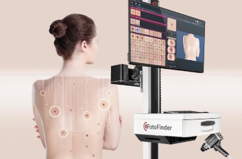
What is digital dermoscopy?
Digital dermoscopy is one of the key modern tools for checking skin lesions. In the hands of a doctor trained in the technique, it shows the structure of a mole or spot in detail, reveals its characteristic dermoscopic features and helps decide what comes next: monitoring, further tests or removal.
Digital dermoscopy is a non-invasive way of examining the skin with special optical equipment. It magnifies the lesion and shows structures that cannot be seen with the naked eye.
Depending on the equipment and settings, the doctor can capture highly magnified images and use additional optical modes for a more detailed assessment.
Dermoscopy is used to assess moles (melanocytic naevi), seborrhoeic keratoses, angiomas, dermatofibromas and other benign or suspicious skin lesions. It also helps diagnose melanoma and non-melanoma skin cancers.
The procedure is painless, involves no surgery and does not damage the skin. For the patient it feels a bit like an ultrasound: the doctor examines the area with a special device, and with digital dermoscopy the image appears on a monitor and can be saved for comparison later.
Who should have their moles and skin lesions checked?
A skin check can be useful for anyone, but how often you need one, and whether digital monitoring makes sense, depends on your personal risk factors, your skin and the results of earlier checks.
Extra attention is needed if you have:
- a large number of moles or atypical moles;
- a personal or family history of melanoma;
- had skin cancer before;
- fair skin that burns easily;
- had a lot of UV exposure over the years, including regular tanning bed use;
- a weakened immune system or other factors that may raise your skin cancer risk;
- new spots, or moles that have started to change.
Lesions that change over time or look clearly different from your other moles deserve a separate look.
See a doctor if you notice:
- a change in a mole's size, shape or colour;
- uneven or blurred edges;
- uneven colouring or new shades appearing;
- bleeding for no obvious reason;
- crusting, an ulcer or discharge;
- itching, pain or any new sensation around the spot;
- a new spot that is changing quickly;
- a sore on the skin that has not healed for a long time.
These signs do not necessarily mean cancer, but they are a reason to have the spot examined by a professional. Do not wait until every symptom on the list appears: suspicious lesions sometimes cause no discomfort at all.
What can dermoscopy detect?
Dermoscopy helps the doctor assess the structure of a lesion and judge whether its features point to a benign or a suspicious process.
In particular, it can help to:
- spot the dermoscopic signs of melanoma;
- suspect basal cell carcinoma and other non-melanoma skin cancers;
- tell different types of benign lesions apart;
- decide whether a lesion needs removing or can be monitored;
- choose the next diagnostic step;
- avoid unnecessary removals in suitable clinical situations;
- record the starting appearance of a lesion for comparison at later visits.
Dermoscopy can also help with some inflammatory and other skin conditions. It is not, however, a universal test for every skin disease.
It is important to understand that dermoscopy is not a final diagnosis. If the doctor suspects melanoma or another skin cancer, removal or a biopsy followed by histology may be needed. In such cases it is histology that confirms the diagnosis.
How does a mole check work?
It starts with a consultation: the doctor asks about your concerns, when the spots appeared and how they have changed, previous diagnoses, family history and risk factors.
The doctor then examines your skin and uses dermoscopy on the lesions that need assessing. If indicated, digital photos are taken so the images can be saved and compared later.
The next step depends on what the doctor finds. Depending on the lesion, it may be:
- Dermoscopic monitoring over time. For lesions that do not need removing straight away but should be kept under watch, dermoscopic images are compared after a set interval.
- Further tests. If the examination cannot reliably establish what the lesion is, other diagnostic methods may be needed.
- Removal with histology. If the lesion looks suspicious, or tissue needs to be examined to make a diagnosis, the doctor chooses the right way to obtain a sample.
Not every mole needs to be removed. But a suspicious lesion should never be removed at home or destroyed with destructive methods before it has been properly assessed.
Digital dermoscopy and monitoring over time
One of the main advantages of digital dermoscopy is that images of individual lesions can be stored and compared with earlier results.
At the first visit, for example, the doctor may identify moles that do not need removing right away but should be watched. Their images are saved, and at the next visit they are compared with new ones.
This comparison helps reveal changes in structure, shape or colour that are easy to miss in an ordinary examination.
Photo documentation and digital monitoring are especially helpful for some patients with many moles or a higher risk of melanoma. Whether you need monitoring, and how often, is decided by the doctor individually.
What equipment is used for mole checks?
Our clinic uses several dermoscopy systems, including the FotoFinder digital imaging system and the DermLite handheld dermatoscope.
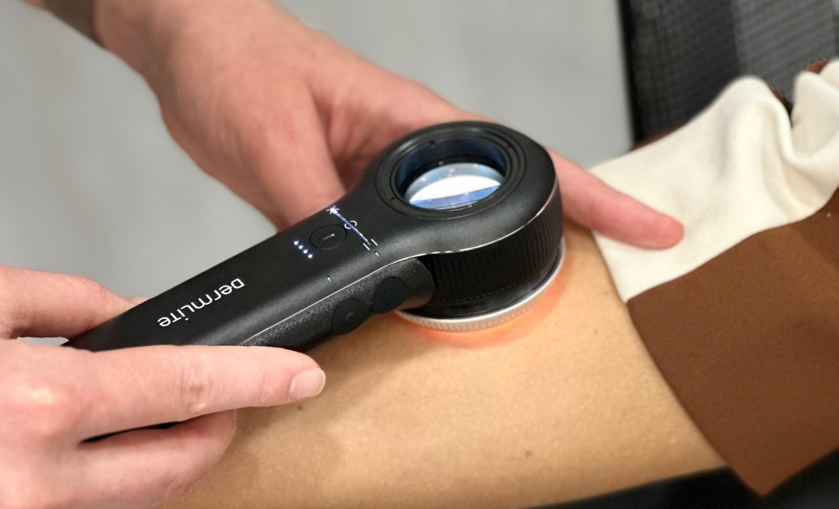
This equipment allows a detailed examination of skin lesions and, when indicated, documenting their appearance for later comparison.
It is worth remembering, though, that a modern device does not guarantee a correct diagnosis on its own. The result depends on the doctor's training, their experience in dermoscopy, how accurately they interpret what they see and the next steps they choose.
The quality of a skin check depends not only on the equipment, but on the specialist who performs it and interprets the results.
How to prepare for dermoscopy
No special preparation is usually needed. Before your visit, however, it helps to:
- avoid foundation or other make-up on the areas to be examined, as it can change how the skin looks;
- if possible, avoid applying creams or ointments to the spot just before the check;
- tell the doctor if a mole has changed, bled, itched or been injured;
- bring the results of earlier examinations, photos or histology reports if you have them.
If only one spot worries you, it can be checked on its own. But if you have never had a skin check before, the doctor may suggest examining other areas too, so that changes elsewhere are not missed.
Why you should not put off a mole check
Skin cancer does not always cause pain or other obvious symptoms. Early changes can be subtle, so it matters to pay attention not only to how you feel, but also to how your skin looks.
How often to have checks should be decided individually, based on your risk factors and earlier results. There is no single interval that suits everyone. Children and adults also need different approaches depending on age, history and clinical situation.
If you have never had dermoscopy, have not had your moles checked for a long time or have noticed changes in your skin, it is worth booking a consultation. Do not wait until a spot starts to hurt or bleed.
Remember: the aim of a check is not to remove as many moles as possible, but to catch suspicious changes in time and to work out which lesions need treatment and which can safely be watched.
Mole and skin lesion checks in Kharkiv
If a mole is bothering you, a new spot has appeared or you would like a preventive skin check, see a doctor who specialises in diagnosing skin lesions.
At the consultation the doctor can examine the lesion with dermoscopy, decide whether digital monitoring is needed and, if indicated, plan further tests or treatment.
Book a mole and skin lesion check in Kharkiv. A timely examination helps catch suspicious changes and choose a well-grounded plan for what to do next.
Get a Consultation
Sources and professional guidelines
- European consensus-based interdisciplinary guideline for melanoma. Part 1: Diagnostics, Update 2024. European Journal of Cancer, 2025
- Russo T. et al. Indications for Digital Monitoring of Patients With Multiple Nevi: Recommendations from the International Dermoscopy Society. Dermatology Practical & Conceptual, 2022
- NICE. Melanoma: assessment and management. Recommendations
- ESMO Clinical Practice Guideline: Cutaneous melanoma, diagnosis, treatment and follow-up
About the author
Nazar V. Tsalin, MD, dermato-oncologist and surgical oncologist.
Focus: diagnosis and surgical treatment of skin lesions, dermoscopy, early detection of skin cancer.