
Mole Removal in Kharkiv: When It's Needed, How It's Done and Whether You Need Histology
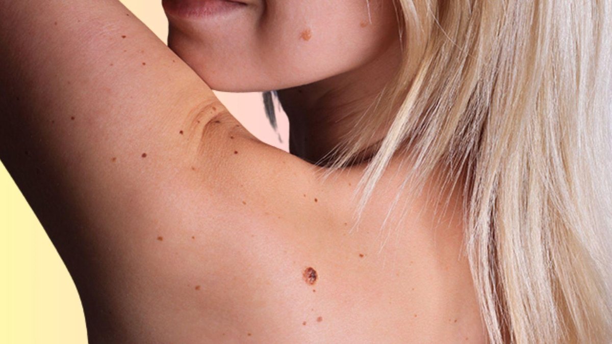
Almost everyone has moles. Some stay the same for years, others slowly grow, change shape or become more noticeable. Sometimes they get nicked while shaving, rub against clothing or simply bother you cosmetically.
That raises fair questions: does the mole need to come off, which method is best, will it scar, and is histology really necessary?
Mole removal in Kharkiv should start with a doctor's consultation and a proper diagnosis. Not every lesion needs removing, but before any procedure it is important to establish what it is, assess its dermoscopic features and choose a method that fits the clinical situation.
As a dermato-oncologist, I want to stress one point: with skin lesions, what matters is not only how a lesion is removed, but whether an accurate diagnosis is still possible afterwards.
What is a mole and why does it appear?
A mole, or melanocytic naevus, is a benign skin growth made up of cells of the melanocytic line.
Melanocytes are the cells that produce melanin, the pigment that colours the skin, hair and iris and helps protect the skin from UV radiation. Melanocytic naevi form from local clusters of naevus cells, which are closely related to melanocytes.
Moles vary in colour, shape, size and texture. They can be light brown, dark brown, almost black, and sometimes pinkish, bluish or greyish. Some lie flat with the skin, others are raised above it.
New moles appearing over a lifetime is a common, normal process. Moles can form in childhood, in the teenage years and in adults. In children, their number and size can change as the body grows.
Still, it is important to tell ordinary age-related changes from signs that need checking. In adults, a new pigmented spot or a change in an existing mole can be a reason for dermoscopy, especially when there are other risk factors.
That does not mean every change is a sign of cancer. But if it has suspicious features, it should not be assumed to be normal without an assessment.
Types of moles and when they should be removed
Moles can be flat or raised, small or large, evenly coloured or uneven. Their appearance does not always reliably show what they are.
For example, intradermal naevi often look like raised, skin-coloured or brown bumps. They can change slowly over a lifetime and eventually start to bother a patient cosmetically.
At the same time, not every raised bump is an intradermal naevus. Other benign and malignant growths can look similar. That is why, before removal, it is important to know exactly what kind of lesion we are dealing with.
Reasons for removal fall roughly into two groups: cosmetic and medical.
Cosmetic reasons for removal
Patients often come in about moles they simply don't like the look of. Most often these are raised lesions on the face, neck and other exposed areas.
Another common reason is regular friction or irritation. A mole may sit in the shaving area, under a belt, on the scalp, in the armpit, under a bra strap or on the back, where it is easily injured in everyday life.
In these cases removal can be considered after a doctor's examination.
However, wanting a lesion gone for cosmetic reasons does not remove the need for a diagnostic assessment first. This matters especially if the mole has changed, is unevenly coloured or looks different from the other spots on your skin.
Medical reasons for removal
Medical reasons arise when a lesion has clinical or dermoscopic features that need a clearer diagnosis, or when a condition requiring treatment has been confirmed.
These situations include:
- suspicious changes in the shape, colour or structure of a mole;
- asymmetry and uneven colouring;
- steady growth or other changes over time;
- dermoscopic features that may point to an atypical naevus or melanoma;
- suspicion of another skin cancer.
An atypical naevus is not the same as melanoma. But some atypical lesions need to be removed and sent for histology if, after the examination, the doctor cannot confidently rule out cancer.
In these cases the aim is not just to get rid of the lesion, but to obtain tissue for an accurate diagnosis.
Why dermoscopy comes before mole removal
Dermoscopy is a non-invasive skin examination with a special optical instrument. It lets the doctor see structures inside a lesion that cannot be properly assessed with the naked eye.
During the examination the doctor looks at pigment structures, colour distribution, symmetry, borders, blood vessels and other features. The findings are weighed together with the patient's history and how the lesion has changed over time.
Dermoscopy helps decide what to do next:
- monitor the lesion if it has benign features;
- monitor it over a short interval if it has atypical structures that do not suggest cancer;
- remove it when there is a reason to, and send the tissue for histology.
The ESMO guidelines stress that dermoscopy performed by an experienced doctor improves the accuracy of melanoma diagnosis.
I do not recommend removing a suspicious pigmented lesion with a destructive method without a proper diagnostic assessment first. If melanoma cannot be ruled out, the approach must allow a diagnosis to be made, not simply destroy the visible part of the lesion.
Mole removal methods
The method depends on the type of lesion, its size and location, the dermoscopic picture and the clinical indications. There is no single method that suits every lesion. The doctor's main job is to assess the lesion correctly and choose a method that allows a safe procedure and, where needed, provides tissue for histology.
1. Superficial removal: shave biopsy
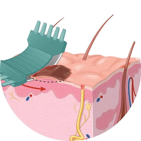
In a shave biopsy the lesion is removed at a set level with a special surgical blade. The tissue is sliced off superficially, hence the name.
This method can be used for certain lesions when it suits the clinical situation. Its key advantage is that the removed tissue can be sent for histology. Still, the doctor must take the possible depth of the lesion into account and perform the procedure so that the sample is good enough for a diagnosis.
If melanoma is suspected, superficial removal must not stand in the way of reliably measuring the depth of the tumour. That is why, where possible, a suspicious melanocytic lesion is removed completely through the full thickness of the skin, with a 1 to 3 mm margin of clinically normal skin. A superficial cut that does not allow the depth of invasion to be assessed properly can complicate further diagnosis and the choice of treatment.
2. Radiowave method
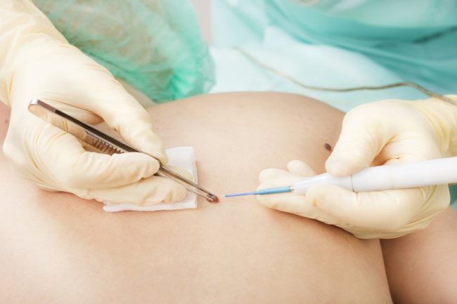
Radiowave instruments can be used for various surgical procedures. It is important to distinguish procedures where the tissue is removed and kept for histology from destructive methods, where the tissue is destroyed.
The radiowave method in itself does not always mean the tissue is destroyed. Whether histology is possible depends on the technique and the condition of the tissue obtained.
If a lesion is completely destroyed without obtaining a usable sample, histology cannot confirm what it was. When cancer is suspected, destructive removal without obtaining tissue for examination is not an acceptable diagnostic approach. In these cases the priority is to obtain tissue that allows an accurate diagnosis.
3. Surgical excision
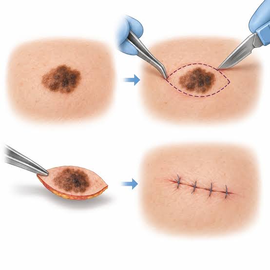
Surgical excision removes the lesion and provides tissue for histology. If melanoma is suspected, the lesion is, where possible, removed completely through the full thickness of the skin with a narrow margin, usually 1 to 3 mm of clinically normal skin.
It is important to understand that this is a diagnostic excision, not the final, definitive operation for a confirmed melanoma. If histology confirms the diagnosis, the extent of further surgery is decided according to the tumour's features and current clinical guidelines.
For large lesions, or lesions in difficult anatomical areas, complete excision at the diagnostic stage may be inappropriate or technically difficult. In these situations the doctor may consider an incisional biopsy, taking a fragment of the lesion for examination. The sample must be representative and contain enough tissue to make the diagnosis and plan what comes next.
This approach is in line with current European and American guidelines on melanoma diagnosis: where possible, a suspicious lesion is removed completely, and in certain clinical situations a limited biopsy is used.
4. Laser removal and cryotherapy
Laser and cryotherapy, which destroys tissue with extreme cold, can be used for certain benign and, very rarely, malignant skin lesions when there are indications. But they are not a universal way to remove moles.
If a lesion has suspicious features, destroying the tissue with a laser, cold or another destructive method can take away the chance of a full histological examination.
That is exactly why European and American melanoma guidelines insist on a proper diagnostic assessment of suspicious lesions and warn against destructive methods while melanoma has not been ruled out. This includes laser ablation, cryotherapy and topical products that destroy tissue.
The right order is to assess the lesion and work out what it most likely is first, and only then choose the method. If a lesion has suspicious features, the priority is a diagnostic approach that allows the tissue to be examined.
Why I insist on histology
Histology is the examination of tissue under a microscope, which shows what a lesion actually is. For suspicious lesions it can be the decisive step in the diagnosis.
The pathologist examines the structure of the tissue and, if cancer is found, describes the features that matter for further treatment. With melanoma, for example, the tumour type, Breslow thickness, ulceration and other morphological and prognostic parameters need to be established.
These findings guide what happens next: whether a repeat, wider excision is needed, how extensive surgery should be and whether further tests are required.
If a suspicious lesion is simply destroyed, leaving no usable tissue, an accurate diagnosis may be impossible. That can make the disease harder to assess, delay the right treatment and limit further planning.
At the same time, histology is not automatically required after every procedure on every typical benign lesion. Whether it is needed depends on the clinical picture, the dermoscopy findings and the method chosen. But if melanoma or another skin cancer is suspected, or a lesion has unusual features, the possibility of a proper histological examination is essential.
Why price alone is a poor reason to choose
The cost of the procedure is an important practical factor, but it should not be the only one.
Sometimes patients choose the quickest or cheapest removal without asking whether the lesion will be examined beforehand and whether tissue will be kept for histology.
In my view, the key question is not how many minutes the procedure takes or which device is used. What matters is whether the indications are right, whether the method suits the lesion and whether the chance to make a diagnosis will be lost.
I advise patients to see specialists with the right medical training, qualifications and experience with skin lesions. Don't hesitate to ask about the doctor's training, their specialty, the removal method and whether histology is possible.
With suspicious lesions, the wrong method can delay the diagnosis. That is why it is worth choosing not just a procedure, but a clinic and a specialist who can provide the full diagnostic process.
How mole removal is done
In most cases removal is done under local anaesthesia. After the anaesthetic injection the area goes numb, so the procedure itself is comfortable.
You may feel touch or pressure, but no pain. If you do feel pain during the procedure, tell the doctor.
How long it takes depends on the size of the lesion, where it is and the method chosen. A simple removal of a small lesion can take just a few minutes, while more complex surgery takes longer.
Afterwards the doctor explains how to care for the wound, when to come back for a check and whether stitches need removing. If the tissue was sent for histology, you need to get the result and discuss it with your doctor.
Will removal leave a scar?
Any procedure that breaks the skin can leave a scar. How it looks depends on the size and depth of the removal, its location, how the wound is closed and how your skin heals.
No one can guarantee removal without any trace. But careful planning and good aftercare help reduce the risk of an unwanted cosmetic result.
If cancer is suspected, the priority is the right diagnosis and treatment. The cosmetic result matters, but it should never be the reason to choose a method that rules out a diagnosis.
How often should moles be checked?
There is no single check-up schedule that suits everyone. How often depends on risk factors, the number and type of moles, your personal and family history and the results of earlier examinations.
For a healthy person with no relevant history, once a year is the usual recommendation.
People with many moles, atypical naevi, a past melanoma or other significant risk factors may be advised to have regular monitoring with dermoscopy or mole mapping.
If a mole starts changing, bleeding or shows other suspicious signs, don't wait for your next scheduled check.
Children also need check-ups when there is a reason, but changes in moles in childhood are judged with age and the clinical context in mind. Not every mole that grows in a child is a sign of cancer.
Mole removal in Kharkiv: who to see
If a mole worries you, keeps getting injured, has changed or you would like it removed for cosmetic reasons, start with a consultation with a doctor who specialises in diagnosing skin lesions.
At the consultation the lesion needs to be assessed, dermoscopy performed, the need for removal decided and a method chosen that fits the clinical situation.
If melanoma or another skin cancer is suspected, the priority is the right diagnostic procedure followed by examination of the tissue. If the lesion is benign, the doctor will help you weigh the removal options, the expected result and the risk of scarring.
Don't choose mole removal on price alone or on a promise that no trace will be left. Choose a specialist who understands skin lesions and can provide a proper diagnosis.
Your health is worth more than any procedure. A timely examination helps you avoid unnecessary procedures and not miss a condition that needs treatment.
Get a Consultation
Scientific sources
- Amaral T. et al. Cutaneous melanoma: ESMO Clinical Practice Guideline for diagnosis, treatment and follow-up. Annals of Oncology. Guideline update, November 2024. Full text in PubMed Central.
- Garbe C. et al. European consensus-based interdisciplinary guideline for melanoma. Part 2: Treatment, Update 2024. European Journal of Cancer, 2025
- National Comprehensive Cancer Network (NCCN). Melanoma: Cutaneous, Clinical Practice Guidelines in Oncology. Guidance on biopsy of suspicious pigmented lesions.
- National Cancer Institute. Common Moles, Dysplastic Nevi, and Risk of Melanoma. Information on moles, risk factors and assessing changes in moles.
- American Academy of Dermatology. Moles: Diagnosis and treatment. Information on clinical assessment and removal of moles.
About the author
Nazar V. Tsalin, MD, dermato-oncologist and surgical oncologist.
Focus: diagnosis and surgical treatment of skin lesions, dermoscopy, early detection of skin cancer.