
Kharkiv's First Dedicated Mohs Surgery Centre: Modern Skin Cancer Treatment with Margin Control
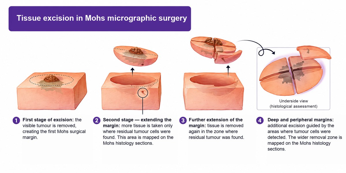
Mohs surgery is a specialised way of treating skin cancer surgically. The tumour is removed in stages, and after each stage the surgical margins are checked under a microscope.
The method was developed by the American surgeon Frederic Edward Mohs in the 1930s. Since then it has evolved a great deal and become one of the most precise surgical approaches to non-melanoma skin cancer.
Today Mohs micrographic surgery is recommended by leading American and European professional societies for certain types of basal cell and squamous cell carcinoma — above all high-risk tumours, recurrent tumours and lesions in anatomically delicate areas.
What sets the method apart is that every stage of removal is checked histologically. This shows whether any tumour cells remain in the surrounding tissue, and if they do, only that affected area is removed next.
The result: the tumour can be removed completely while as much healthy tissue as possible is preserved. The surgical defect stays smaller, and reconstruction afterwards is easier.
This is exactly how we work at our Mohs Surgery Centre in Kharkiv. The goal is not just to remove the tumour you can see, but to check its borders fully under the microscope, keep the treatment oncologically safe and spare as much healthy tissue as possible.
In clinical practice, the local recurrence rate after Mohs surgery is below 1%.
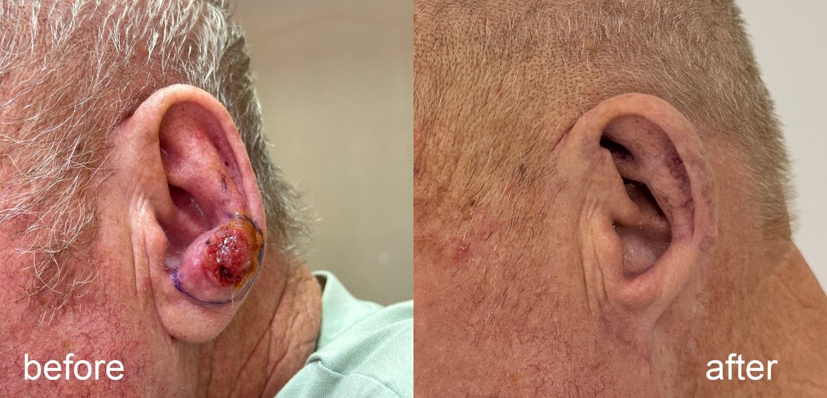
The patient before surgery and at the follow-up visit 3 months later.
The operation is usually done under local anaesthetic as an outpatient procedure. In most cases the patient goes home the same day.
Who is Mohs surgery for?
The main indications for Mohs surgery are basal cell carcinoma (BCC) and squamous cell carcinoma (SCC) of the skin.
Whether the method is right depends on the histological type of the tumour, its size, location, clinical borders, depth of invasion, any high-risk features and the patient's general health.
Mohs surgery is especially worth considering when:
- the clinical borders of the tumour are unclear or poorly defined;
- a BCC or SCC has come back;
- a previous removal was incomplete;
- the tumour has reappeared after earlier treatment;
- the histological subtype is aggressive;
- there is perineural invasion — tumour cells spreading along nerves;
- an SCC shows vascular or lymphatic invasion;
- the tumour has grown deep into the subcutaneous tissue or deeper structures;
- the patient is immunosuppressed, for example after an organ transplant or on immunosuppressive therapy;
- the tumour has developed in an area treated with radiotherapy before;
- preserving as much healthy tissue as possible is essential.
One of the most dangerous situations of all is a tumour that someone has already tried to remove with destructive methods — laser, cryotherapy, or at home with harsh topical remedies such as celandine-based preparations. If tumour tissue remains or the lesion comes back after this, it needs a fresh diagnostic assessment and a new treatment plan.
In such cases another superficial removal is not enough: the key is to establish the histological nature of the lesion and assess its true borders.
Which areas need particular care?
One of the main strengths of Mohs surgery is that it spares as much healthy tissue as possible where every millimetre matters, anatomically and functionally.
Precise mapping of the tumour borders is especially important on:
- the central face;
- the nose and the area around it down to the lips;
- the eyelids, eyebrows and the skin around the eyes;
- the lips;
- the cheeks and forehead;
- the chin and jawline;
- the ears and the skin in front of them;
- the temples;
- the scalp;
- the neck;
- the hands and feet;
- areas of the legs where a functional or cosmetic defect is likely;
- the external genitals.
Location is not the only factor in choosing the method. Even a small tumour on the eyelid or nose may need micrographic surgery because the structures around it must be preserved. On the other hand, for some lesions on the trunk or limbs a standard surgical excision may be enough — though not always.
Is Mohs surgery used for other tumours?
Yes. Besides basal cell and squamous cell carcinoma, micrographic surgery can be used for some rare skin cancers, including:
- dermatofibrosarcoma protuberans (DFSP);
- sebaceous carcinoma;
- microcystic adnexal carcinoma;
- certain other tumours of the skin appendages.
For these tumours the decision is made case by case, based on the histological diagnosis, location, pattern of spread and how well the margins can be checked under the microscope.
Mohs surgery is not a universal method for every skin cancer. For melanoma in particular, it needs specific indications, special histological techniques and the right clinical experience.
How is Mohs surgery better than standard excision?
In a standard surgical excision, the tumour is cut out with a minimal margin around its visible borders. The tissue is sent for histology, but with the standard technique the margins are usually assessed on selected sections only.
In Mohs surgery the tissue is processed and oriented in a special way, so the entire surface of the peripheral and deep margins of the specimen can be examined.
If tumour cells remain, the surgeon knows exactly where they are and removes more tissue only in that spot.
The main advantages of the method:
- Microscopic margin control. The peripheral and deep borders of the tissue are assessed during the operation itself.
- Maximum preservation of healthy tissue. Further excision is targeted, rather than widening the whole surgical field without need.
- Control of microscopic tumour spread. Tumour cells beyond the visible clinical borders can be found.
- A smaller surgical defect. The healthy tissue that is spared makes reconstruction easier.
- High local control. The method is especially valuable for high-risk and recurrent tumours.
In short, Mohs surgery brings together two goals that matter: removing the tumour completely and keeping as much healthy tissue as possible.
How is Mohs surgery done?
Mohs surgery is usually an outpatient procedure under local anaesthetic. The patient stays awake, and only the area being operated on is numbed. The patient feels no pain during the operation. In most cases no hospital stay is needed.
Before the procedure the doctor reviews the patient's general health, other conditions and any medicines they take, especially anticoagulants. The histological diagnosis is confirmed and the extent of surgery is planned.
Whether lab tests or other examinations are needed is decided individually. There is no single list of tests that every patient must have before outpatient Mohs surgery.
The number of stages depends on the location and size of the tumour, its morphological type, how far it extends beyond the visible margins and the patient's individual features. In practice 2–3 stages are often needed, followed by reconstruction of the surgical defect. Sometimes a single stage is enough.
Stage one — removing the tumour
Once the local anaesthetic has taken effect, the surgeon removes the visible tumour using the Mohs technique.
The tissue goes for specialised histological processing. It is oriented, marked with special dyes and divided into sections according to a map of the specimen.
Under the microscope, the pathologist examines the peripheral and deep margins of the removed tissue. Because the specimen is oriented correctly, it is possible to see exactly where any remaining tumour cells are.
While the lab processes and examines the tissue, the wound is covered with a temporary dressing. The patient waits for the result under the care of the medical team.
Stage two — targeted removal of remaining tumour cells
If tumour cells are found under the microscope, their position is marked on the map of the surgical field.
Using this map, the surgeon pinpoints the exact area where the rest of the tumour has to be removed. The next stage is carried out only in the zone where histology has confirmed residual tumour.
The newly excised tissue again goes through histological processing and microscopic assessment.
This cycle is repeated as many times as needed to reach the main goal: no tumour cells in the examined peripheral and deep margins of the specimen.
At each stage, only the piece of tissue where residual tumour was found is removed. This preserves as much healthy skin and surrounding structure as possible.
Final stage — reconstructing the defect
Once histology confirms that the tumour has been removed completely, the surgeon closes the surgical defect.
The way it is reconstructed depends on the size and depth of the wound, its location, the state of the surrounding tissue and what will give the best functional and cosmetic result.
Depending on the clinical situation, the options include:
- linear (straight-line) closure of the wound;
- a local flap, moving nearby skin to cover the defect;
- a skin graft;
- healing by secondary intention, if the location and type of defect allow it.
Not every defect can simply be stitched in a straight line. Sometimes a proper reconstruction needs additional incisions and repositioning of the surrounding tissue.
That is why reconstruction is planned as a step of its own. Its aim is not only to close the wound but to preserve function as fully as possible and achieve the best cosmetic result.
How long does Mohs surgery take?
It varies from patient to patient. The length depends on the size and location of the tumour, the number of stages needed, the time histological processing takes and how complex the reconstruction is.
It is best to set aside the whole day, because the number of stages cannot be predicted in advance. Even a small lesion sometimes extends microscopically beyond its visible borders.
Do you need to stay in hospital after Mohs surgery?
In most cases Mohs surgery is done as an outpatient procedure. Once the operation is over, and if there are no medical reasons against it, the patient can leave the clinic and recover at home.
After the procedure patients can usually move about and look after themselves. When it is safe to drive, go back to work or do housework is decided individually, depending on the extent of surgery, where the wound is and the surgeon's advice.
A hospital stay may be needed in exceptional cases — for example, with complex coexisting conditions, extensive reconstruction, or when general anaesthesia and post-operative monitoring are required.
Will Mohs surgery leave a scar?
Yes. Like any surgery, Mohs surgery leaves a scar. It is important to be clear about this: it is an operation.
But one of the key strengths of the method is that it preserves as much healthy tissue as possible. This keeps the surgical defect as small as complete removal of the tumour allows.
What the scar will look like depends on several factors:
- the size and depth of the final surgical defect;
- its location on the body;
- the reconstruction technique chosen;
- the patient's age;
- how the patient's skin heals;
- other conditions and factors that affect healing.
The final look of a scar cannot always be predicted precisely. It depends not only on the stitching technique but also on how each person's tissue responds to surgery.
So when we plan an operation, we think not only about removing the tumour completely but also about the reconstruction that follows, taking the anatomy and appearance of the area into account.
What is recovery like?
After the procedure the patient receives personal instructions on wound care, dressings, physical activity and follow-up visits.
As a rule, you will need to:
- follow the instructions for caring for the dressing and the wound;
- come to follow-up visits and dressing changes on time;
- avoid putting too much tension on the tissue around the operated area;
- cut back on strenuous exercise for a while, if the surgeon advises it;
- watch for signs of possible complications and tell your doctor about them.
The first follow-up visit or dressing change usually takes place within a few days of surgery. The exact timing depends on the type of reconstruction and how the wound looks.
Stitches are usually removed after about 8–12 days, though this can vary with the location of the surgery and how the wound heals.
After that, the patient keeps caring for the scar as the doctor advises. A scar takes much longer to mature than the wound takes to heal, so the final cosmetic result is judged over time, not right after the stitches come out.
The main purpose of aftercare is proper healing, a lower risk of complications and the best possible functional and cosmetic result.
Why choose a dedicated Mohs Surgery Centre?
Mohs surgery is not simply cutting out a lesion with a surgical instrument. It is a complex process: the surgeon and the pathologist working in step, specialised histological processing, correct orientation of every specimen and the ability to carry out extra stages of removal based on what the microscope shows.
The accuracy of margin control and the outcome of treatment depend on the quality of each of these steps.
At our Centre we pay attention to every part of the treatment:
- assessing the lesion beforehand, including its histological features;
- selecting patients for Mohs surgery in line with international guidelines;
- precise surgical removal of the tumour;
- microscopic control of the peripheral and deep margins;
- planning the reconstruction of the defect;
- aftercare and long-term follow-up.
Our aim is not just to remove the tumour, but to provide sound oncological control, preserve as much healthy tissue as possible and achieve the best functional and cosmetic result.
If you have been diagnosed with basal cell or squamous cell carcinoma — especially on the face, nose, eyelids or ears, or anywhere else — talk to a dermato-oncologist about whether Mohs surgery is right for you.
The Mohs Surgery Centre in Kharkiv: a specialised approach to skin cancer treatment with microscopic control of the surgical margins.
Book a consultation to review your treatment options and choose the best approach for your case.
Diagnosis and treatment at the Institute
Get a consultation
References
- Murray C. et al. Patient Indications for Mohs Micrographic Surgery: A Clinical Practice Guideline. Current Oncology. 2019. PubMed | Full text.
- Peris K. et al. European Consensus-Based Interdisciplinary Guideline for Diagnosis and Treatment of Basal Cell Carcinoma — Update 2023. European Journal of Cancer. 2023. Full text.
- Lang B. M. et al. S2k Guideline Basal Cell Carcinoma of the Skin (Update 2023). Journal der Deutschen Dermatologischen Gesellschaft. 2024. PubMed Central.
- Alam M. et al. Guidelines of Care for the Management of Cutaneous Squamous Cell Carcinoma. Journal of the American Academy of Dermatology. 2018. PubMed | Full text.
- Connolly S. M. et al. Mohs Micrographic Surgery: A Review of Indications, Technique, Outcomes, and Considerations. Anais Brasileiros de Dermatologia. 2021. PubMed | Full text.
- Lacerda P. N. et al. Recurrence Rate of Basal Cell Carcinoma Among Different Micrographic Surgery Techniques: Systematic Review with Meta-Analysis. Journal of the European Academy of Dermatology and Venereology. 2022. PubMed.
- NCCN Clinical Practice Guidelines in Oncology: Basal Cell Skin Cancer. National Comprehensive Cancer Network (NCCN). Official NCCN resource.
- NCCN Clinical Practice Guidelines in Oncology: Squamous Cell Skin Cancer. National Comprehensive Cancer Network (NCCN). Official NCCN resource.
About the author
Nazar V. Tsalin, MD, dermato-oncologist and surgical oncologist.
Areas of focus: diagnosis and surgical treatment of skin lesions, dermoscopy, early detection of skin cancer, Mohs surgery.